3UAU | pdb_00003uau

Crystal structure of the lipoprotein JlpA


Experimental Data Snapshot

  • Method: X-RAY DIFFRACTION
  • Resolution: 2.70 Å
  • R-Value Free: 
    0.272 (Depositor), 0.273 (DCC) 
  • R-Value Work: 
    0.242 (Depositor), 0.243 (DCC) 
  • R-Value Observed: 
    0.243 (Depositor) 

wwPDB Validation 3D Report Full Report

Validation slider image for 3UAU

This is version 1.1 of the entry. See complete history

Literature

Crystal structure of JlpA, a surface-exposed lipoprotein adhesin of Campylobacter jejuni.

Kawai, F.Paek, S.Choi, K.J.Prouty, M.Kanipes, M.I.Guerry, P.Yeo, H.J.

(2012) J Struct Biol 177: 583-588

  • DOI: https://doi.org/10.1016/j.jsb.2012.01.001
  • Primary Citation Related Structures: 
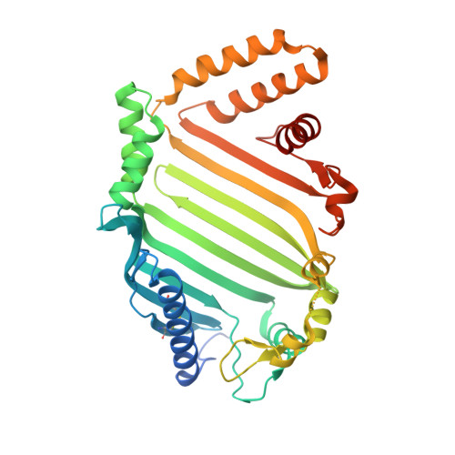
    3UAU

  • PubMed Abstract: 

    The Campylobacter jejuni JlpA protein is a surface-exposed lipoprotein that was discovered as an adhesin promoting interaction with host epithelium cells, an early critical step in the pathogenesis of C. jejuni disease. Increasing evidence ascertained that JlpA is antigenic, indicating a role of JlpA in immune response during the infectious process. Here, we report the crystal structure of JlpA at 2.7Å resolution, revealing a catcher's mitt shaped unclosed half β-barrel. Although the apparent architecture of JlpA is somewhat reminiscent of other bacterial lipoproteins such as LolB, the topology of JlpA is unique among the bacterial surface proteins reported to date and therefore JlpA represents a novel bacterial cell surface lipoprotein. The concave face of the structure results in an unusually large hydrophobic basin with a localized acidic pocket, suggesting a possibility that JlpA may accommodate multiple ligands. Therefore, the structure provides framework for determining the molecular function of JlpA and new strategies for the rational design of small molecule inhibitors efficiently targeting JlpA.


  • Organizational Affiliation
    • Department of Biology and Biochemistry, University of Houston, Houston, TX 77204, USA.

Macromolecule Content 

  • Total Structure Weight: 86.77 kDa 
  • Atom Count: 5,510 
  • Modeled Residue Count: 685 
  • Deposited Residue Count: 758 
  • Unique protein chains: 1

Macromolecules

Find similar proteins by:|  3D Structure
Entity ID: 1
MoleculeChains  Sequence LengthOrganismDetailsImage
Surface-exposed lipoprotein
A, B
379Campylobacter jejuni subsp. jejuni 81-176Mutation(s): 0 
Gene Names: CJJ81176_1002jlpA
UniProt
Find proteins for A0A0H3P9U7 (Campylobacter jejuni subsp. jejuni serotype O:23/36 (strain 81-176))
Explore A0A0H3P9U7 
Go to UniProtKB:  A0A0H3P9U7
Entity Groups
Sequence Clusters30% Identity50% Identity70% Identity90% Identity95% Identity100% Identity
UniProt GroupA0A0H3P9U7
Sequence Annotations
Expand
Reference Sequence

Experimental Data & Validation

Experimental Data

  • Method: X-RAY DIFFRACTION
  • Resolution: 2.70 Å
  • R-Value Free:  0.272 (Depositor), 0.273 (DCC) 
  • R-Value Work:  0.242 (Depositor), 0.243 (DCC) 
  • R-Value Observed: 0.243 (Depositor) 
Space Group: P 41 21 2
Unit Cell:
Length ( Å )Angle ( ˚ )
a = 111.632α = 90
b = 111.632β = 90
c = 170.369γ = 90
Software Package:
Software NamePurpose
HKL-3000data collection
SHARPphasing
REFMACrefinement
HKL-3000data reduction
SCALEPACKdata scaling

Structure Validation

View Full Validation Report


Entry History 

Deposition Data

Revision History  (Full details and data files)

  • Version 1.0: 2012-07-25
    Type: Initial release
  • Version 1.1: 2024-10-16
    Changes: Data collection, Database references, Structure summary