3UA7 | pdb_00003ua7

Crystal Structure of the Human Fyn SH3 domain in complex with a peptide from the Hepatitis C virus NS5A-protein


Experimental Data Snapshot

  • Method: X-RAY DIFFRACTION
  • Resolution: 1.50 Å
  • R-Value Free: 
    0.231 (Depositor), 0.233 (DCC) 
  • R-Value Work: 
    0.185 (Depositor), 0.187 (DCC) 
  • R-Value Observed: 
    0.190 (Depositor) 

Starting Model: experimental
View more details

wwPDB Validation 3D Report Full Report

Validation slider image for 3UA7

This is version 1.3 of the entry. See complete history

Literature

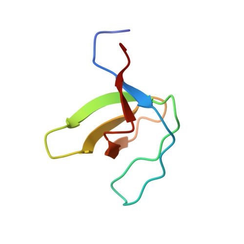
The promiscuous binding of the Fyn SH3 domain to a peptide from the NS5A protein.

Martin-Garcia, J.M.Luque, I.Ruiz-Sanz, J.Camara-Artigas, A.

(2012) Acta Crystallogr D Biol Crystallogr 68: 1030-1040

  • DOI: https://doi.org/10.1107/S0907444912019798
  • Primary Citation Related Structures: 
    3UA6, 3UA7

  • PubMed Abstract: 

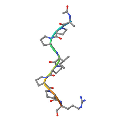
    The hepatitis C virus nonstructural 5A (NS5A) protein is a large zinc-binding phosphoprotein that plays an important role in viral RNA replication and is involved in altering signal transduction pathways in the host cell. This protein interacts with Fyn tyrosine kinase in vivo and regulates its kinase activity. The 1.5 Å resolution crystal structure of a complex between the SH3 domain of the Fyn tyrosine kinase and the C-terminal proline-rich motif of the NS5A-derived peptide APPIPPPRRKR has been solved. Crystals were obtained in the presence of ZnCl(2) and belonged to the tetragonal space group P4(1)2(1)2. The asymmetric unit is composed of four SH3 domains and two NS5A peptide molecules; only three of the domain molecules contain a bound peptide, while the fourth molecule seems to correspond to a free form of the domain. Additionally, two of the SH3 domains are bound to the same peptide chain and form a ternary complex. The proline-rich motif present in the NS5A protein seems to be important for RNA replication and virus assembly, and the promiscuous interaction of the Fyn SH3 domain with the NS5A C-terminal proline-rich peptide found in this crystallographic structure may be important in the virus infection cycle.


  • Organizational Affiliation
    • Department of Physical Chemistry, Biochemistry and Inorganic Chemistry, University of Almería, Agrifood Campus of International Excellence (ceiA3), Carretera de Sacramento, 04120 Almería, Spain.

Macromolecule Content 

  • Total Structure Weight: 31.69 kDa 
  • Atom Count: 2,266 
  • Modeled Residue Count: 263 
  • Deposited Residue Count: 280 
  • Unique protein chains: 2

Macromolecules

Find similar proteins by:|  3D Structure
Entity ID: 1
MoleculeChains  Sequence LengthOrganismDetailsImage
Tyrosine-protein kinase Fyn
A, B, C, D
64Homo sapiensMutation(s): 0 
Gene Names: FYN
EC: 2.7.10.2
UniProt & NIH Common Fund Data Resources
Find proteins for P06241 (Homo sapiens)
Explore P06241 
Go to UniProtKB:  P06241
PHAROS:  P06241
GTEx:  ENSG00000010810 
Entity Groups
Sequence Clusters30% Identity50% Identity70% Identity90% Identity95% Identity100% Identity
UniProt GroupP06241
Sequence Annotations
Expand
Reference Sequence
Find similar proteins by:  Sequence   |   3D Structure  
Entity ID: 2
MoleculeChains  Sequence LengthOrganismDetailsImage
Non-structural protein 5A
E, F
12Orthohepacivirus hominisMutation(s): 1 
UniProt
Find proteins for P26663 (Hepatitis C virus genotype 1b (isolate BK))
Explore P26663 
Go to UniProtKB:  P26663
Entity Groups
Sequence Clusters30% Identity50% Identity70% Identity90% Identity95% Identity100% Identity
UniProt GroupP26663
Sequence Annotations
Expand
Reference Sequence

Small Molecules

Ligands 5 Unique
IDChains Name / Formula / InChI Key2D Diagram3D Interactions
GOL

Query on GOL


Download:Ideal Coordinates CCD File
G [auth A]GLYCEROL
C3 H8 O3
PEDCQBHIVMGVHV-UHFFFAOYSA-N
ZN

Query on ZN


Download:Ideal Coordinates CCD File
J [auth C],
L [auth D]
ZINC ION
Zn
PTFCDOFLOPIGGS-UHFFFAOYSA-N
FMT

Query on FMT


Download:Ideal Coordinates CCD File
K [auth C],
M [auth D]
FORMIC ACID
C H2 O2
BDAGIHXWWSANSR-UHFFFAOYSA-N
CL

Query on CL


Download:Ideal Coordinates CCD File
H [auth A]CHLORIDE ION
Cl
VEXZGXHMUGYJMC-UHFFFAOYSA-M
NA

Query on NA


Download:Ideal Coordinates CCD File
I [auth A]SODIUM ION
Na
FKNQFGJONOIPTF-UHFFFAOYSA-N

Experimental Data & Validation

Experimental Data

  • Method: X-RAY DIFFRACTION
  • Resolution: 1.50 Å
  • R-Value Free:  0.231 (Depositor), 0.233 (DCC) 
  • R-Value Work:  0.185 (Depositor), 0.187 (DCC) 
  • R-Value Observed: 0.190 (Depositor) 
Space Group: P 41 21 2
Unit Cell:
Length ( Å )Angle ( ˚ )
a = 51.39α = 90
b = 51.39β = 90
c = 185.59γ = 90
Software Package:
Software NamePurpose
MOSFLMdata reduction
SCALAdata scaling
MOLREPphasing
REFMACrefinement
PDB_EXTRACTdata extraction
ADSCdata collection

Structure Validation

View Full Validation Report


Entry History 

Deposition Data

Revision History  (Full details and data files)

  • Version 1.0: 2012-07-25
    Type: Initial release
  • Version 1.1: 2013-01-23
    Changes: Database references
  • Version 1.2: 2023-09-13
    Changes: Data collection, Database references, Derived calculations, Refinement description
  • Version 1.3: 2024-10-30
    Changes: Structure summary