


Crystal structure of Human Apurinic/Apyridinimic Endonuclease, Ape1 in a new crystal form
Agarwal, R., Naidu, M.D.To be published.
Experimental Data Snapshot
Starting Model: experimental
View more details
wwPDB Validation 3D Report Full Report

Macromolecule Content
Entity ID: 1 | |||||
|---|---|---|---|---|---|
| Molecule | Chains | Sequence Length | Organism | Details | Image |
| DNA-(apurinic or apyrimidinic site) lyase | 318 | Homo sapiens | Mutation(s): 0 Gene Names: APEX1, APE, APE1, APEX, APX, HAP1, REF1 EC: 3.1 (PDB Primary Data), 4.2.99.18 (PDB Primary Data), 3.1.21 (UniProt), 3.1.11.2 (UniProt) | 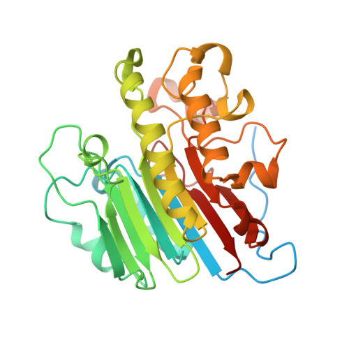 | |
UniProt & NIH Common Fund Data Resources | |||||
PHAROS: P27695 GTEx: ENSG00000100823 | |||||
Entity Groups | |||||
| Sequence Clusters | 30% Identity50% Identity70% Identity90% Identity95% Identity100% Identity | ||||
| UniProt Group | P27695 | ||||
Sequence AnnotationsExpand | |||||
Reference Sequence | |||||
| Ligands 2 Unique | |||||
|---|---|---|---|---|---|
| ID | Chains | Name / Formula / InChI Key | 2D Diagram | 3D Interactions | |
| CL Download:Ideal Coordinates CCD File | H [auth A], J [auth B], L [auth C], P [auth F] | CHLORIDE ION Cl VEXZGXHMUGYJMC-UHFFFAOYSA-M |  | ||
| MG Download:Ideal Coordinates CCD File | G [auth A] I [auth B] K [auth C] M [auth D] N [auth E] | MAGNESIUM ION Mg JLVVSXFLKOJNIY-UHFFFAOYSA-N |  | ||
| Length ( Å ) | Angle ( ˚ ) |
|---|---|
| a = 95.84 | α = 90 |
| b = 97.284 | β = 90.91 |
| c = 132.15 | γ = 90 |
| Software Name | Purpose |
|---|---|
| CBASS | data collection |
| MOLREP | phasing |
| PHASER | phasing |
| REFMAC | refinement |
| HKL-2000 | data reduction |
| HKL-2000 | data scaling |


