CD38 Structure-Based Inhibitor Design Using the N1-Cyclic Inosine 5'-Diphosphate Ribose Template
Moreau, C., Liu, Q., Graeff, R., Wagner, G.K., Thomas, M.P., Swarbrick, J.M., Shuto, S., Lee, H.C., Hao, Q., Potter, B.V.L.(2013) PLoS One 8: e66247-e66247
- PubMed: 23840430 Search on PubMedSearch on PubMed Central
- DOI: https://doi.org/10.1371/journal.pone.0066247
- Primary Citation Related Structures:
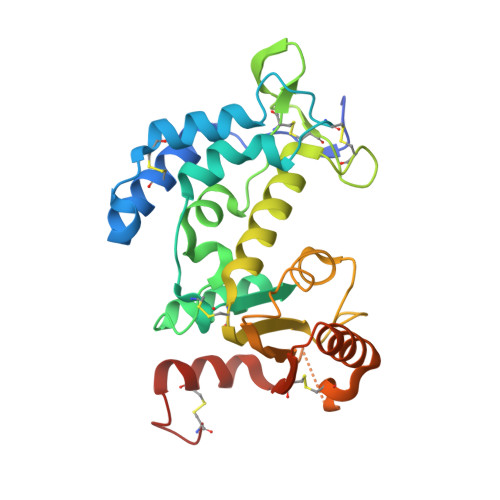
3U4H, 3U4I - PubMed Abstract:
Few inhibitors exist for CD38, a multifunctional enzyme catalyzing the formation and metabolism of the Ca(2+)-mobilizing second messenger cyclic adenosine 5'-diphosphoribose (cADPR). Synthetic, non-hydrolyzable ligands can facilitate structure-based inhibitor design. Molecular docking was used to reproduce the crystallographic binding mode of cyclic inosine 5'-diphosphoribose (N1-cIDPR) with CD38, revealing an exploitable pocket and predicting the potential to introduce an extra hydrogen bond interaction with Asp-155. The purine C-8 position of N1-cIDPR (IC50 276 µM) was extended with an amino or diaminobutane group and the 8-modified compounds were evaluated against CD38-catalyzed cADPR hydrolysis. Crystallography of an 8-amino N1-cIDPR:CD38 complex confirmed the predicted interaction with Asp-155, together with a second H-bond from a realigned Glu-146, rationalizing the improved inhibition (IC50 56 µM). Crystallography of a complex of cyclic ADP-carbocyclic ribose (cADPcR, IC50 129 µM) with CD38 illustrated that Glu-146 hydrogen bonds with the ligand N6-amino group. Both 8-amino N1-cIDPR and cADPcR bind deep in the active site reaching the catalytic residue Glu-226, and mimicking the likely location of cADPR during catalysis. Substantial overlap of the N1-cIDPR "northern" ribose monophosphate and the cADPcR carbocyclic ribose monophosphate regions suggests that this area is crucial for inhibitor design, leading to a new compound series of N1-inosine 5'-monophosphates (N1-IMPs). These small fragments inhibit hydrolysis of cADPR more efficiently than the parent cyclic compounds, with the best in the series demonstrating potent inhibition (IC50 = 7.6 µM). The lower molecular weight and relative simplicity of these compounds compared to cADPR make them attractive as a starting point for further inhibitor design.
- Wolfson Laboratory of Medicinal Chemistry, Department of Pharmacy and Pharmacology, University of Bath, Bath, United Kingdom.
Organizational Affiliation: