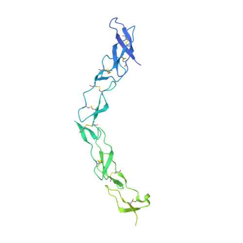
3U3S | pdb_00003u3s

The S-SAD phased crystal structure of the ecto-domain of Death Receptor 6 (DR6)


Experimental Data Snapshot

  • Method: X-RAY DIFFRACTION
  • Resolution: 2.70 Å
  • R-Value Free: 
    0.260 (Depositor), 0.261 (DCC) 
  • R-Value Work: 
    0.197 (Depositor), 0.196 (DCC) 
  • R-Value Observed: 
    0.199 (Depositor) 

wwPDB Validation 3D Report Full Report

Validation slider image for 3U3S

This is version 1.2 of the entry. See complete history. 

Literature

S-SAD phasing study of death receptor 6 and its solution conformation revealed by SAXS.

Ru, H., Zhao, L., Ding, W., Jiao, L., Shaw, N., Liang, W., Zhang, L., Hung, L.W., Matsugaki, N., Wakatsuki, S., Liu, Z.J.

(2012) Acta Crystallogr D Biol Crystallogr 68: 521-530

  • DOI: https://doi.org/10.1107/S0907444912004490
  • Primary Citation Related Structures: 
    3U3P, 3U3Q, 3U3S, 3U3T, 3U3V

  • PubMed Abstract: 

    A subset of tumour necrosis factor receptor (TNFR) superfamily members contain death domains in their cytoplasmic tails. Death receptor 6 (DR6) is one such member and can trigger apoptosis upon the binding of a ligand by its cysteine-rich domains (CRDs). The crystal structure of the ectodomain (amino acids 1-348) of human death receptor 6 (DR6) encompassing the CRD region was phased using the anomalous signal from S atoms. In order to explore the feasibility of S-SAD phasing at longer wavelengths (beyond 2.5 Å), a comparative study was performed on data collected at wavelengths of 2.0 and 2.7 Å. In spite of sub-optimal experimental conditions, the 2.7 Å wavelength used for data collection showed potential for S-SAD phasing. The results showed that the R(ano)/R(p.i.m.) ratio is a good indicator for monitoring the anomalous data quality when the anomalous signal is relatively strong, while d''/sig(d'') calculated by SHELXC is a more sensitive and stable indicator applicable for grading a wider range of anomalous data qualities. The use of the `parameter-space screening method' for S-SAD phasing resulted in solutions for data sets that failed during manual attempts. SAXS measurements on the ectodomain suggested that a dimer defines the minimal physical unit of an unliganded DR6 molecule in solution.


  • Organizational Affiliation: 
    • National Laboratory of Biomacromolecules, Institute of Biophysics, Chinese Academy of Sciences, Beijing 100101, People's Republic of China.

Macromolecule Content 

  • Total Structure Weight: 34.21 kDa 
  • Atom Count: 1,375 
  • Modeled Residue Count: 164 
  • Deposited Residue Count: 313 
  • Unique protein chains: 1

Macromolecules

Find similar proteins by:|  3D Structure
Entity ID: 1
MoleculeChains  Sequence LengthOrganismDetailsImage
Tumor necrosis factor receptor superfamily member 21313Homo sapiensMutation(s): 0 
Gene Names: TNFRSF21, DR6, UNQ437/PRO868
UniProt & NIH Common Fund Data Resources
Find proteins for O75509 (Homo sapiens)
Explore O75509 
Go to UniProtKB:  O75509
PHAROS:  O75509
GTEx:  ENSG00000146072 
Entity Groups
Sequence Clusters30% Identity50% Identity70% Identity90% Identity95% Identity100% Identity
UniProt GroupO75509
Sequence Annotations
Expand
Reference Sequence

Experimental Data & Validation

Experimental Data

  • Method: X-RAY DIFFRACTION
  • Resolution: 2.70 Å
  • R-Value Free:  0.260 (Depositor), 0.261 (DCC) 
  • R-Value Work:  0.197 (Depositor), 0.196 (DCC) 
  • R-Value Observed: 0.199 (Depositor) 
Space Group: P 61 2 2
Unit Cell:
Length ( Å )Angle ( ˚ )
a = 77.674α = 90
b = 77.674β = 90
c = 186.8γ = 120
Software Package:
Software NamePurpose
HKL-2000data collection
SHELXSphasing
REFMACrefinement
HKL-2000data reduction
HKL-2000data scaling

Structure Validation

View Full Validation Report


Entry History 

Deposition Data

Revision History  (Full details and data files)

  • Version 1.0: 2012-05-02
    Type: Initial release
  • Version 1.1: 2013-07-10
    Changes: Database references
  • Version 1.2: 2024-10-16
    Changes: Data collection, Database references, Structure summary