3U2Z | pdb_00003u2z

Activator-Bound Structure of Human Pyruvate Kinase M2


Experimental Data Snapshot

  • Method: X-RAY DIFFRACTION
  • Resolution: 2.10 Å
  • R-Value Free: 
    0.231 (Depositor), 0.233 (DCC) 
  • R-Value Work: 
    0.190 (Depositor), 0.197 (DCC) 
  • R-Value Observed: 
    0.191 (Depositor) 

Starting Model: experimental
View more details

wwPDB Validation 3D Report Full Report

Validation slider image for 3U2Z

Ligand Structure Quality Assessment 


This is version 1.4 of the entry. See complete history

Literature

Pyruvate kinase M2 activators promote tetramer formation and suppress tumorigenesis.

Anastasiou, D.Yu, Y.Israelsen, W.J.Jiang, J.K.Boxer, M.B.Hong, B.S.Tempel, W.Dimov, S.Shen, M.Jha, A.Yang, H.Mattaini, K.R.Metallo, C.M.Fiske, B.P.Courtney, K.D.Malstrom, S.Khan, T.M.Kung, C.Skoumbourdis, A.P.Veith, H.Southall, N.Walsh, M.J.Brimacombe, K.R.Leister, W.Lunt, S.Y.Johnson, Z.R.Yen, K.E.Kunii, K.Davidson, S.M.Christofk, H.R.Austin, C.P.Inglese, J.Harris, M.H.Asara, J.M.Stephanopoulos, G.Salituro, F.G.Jin, S.Dang, L.Auld, D.S.Park, H.W.Cantley, L.C.Thomas, C.J.Vander Heiden, M.G.

(2012) Nat Chem Biol 8: 839-847

  • DOI: https://doi.org/10.1038/nchembio.1060
  • Primary Citation Related Structures: 
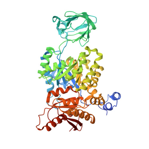
    3ME3, 3U2Z

  • PubMed Abstract: 

    Cancer cells engage in a metabolic program to enhance biosynthesis and support cell proliferation. The regulatory properties of pyruvate kinase M2 (PKM2) influence altered glucose metabolism in cancer. The interaction of PKM2 with phosphotyrosine-containing proteins inhibits enzyme activity and increases the availability of glycolytic metabolites to support cell proliferation. This suggests that high pyruvate kinase activity may suppress tumor growth. We show that expression of PKM1, the pyruvate kinase isoform with high constitutive activity, or exposure to published small-molecule PKM2 activators inhibits the growth of xenograft tumors. Structural studies reveal that small-molecule activators bind PKM2 at the subunit interaction interface, a site that is distinct from that of the endogenous activator fructose-1,6-bisphosphate (FBP). However, unlike FBP, binding of activators to PKM2 promotes a constitutively active enzyme state that is resistant to inhibition by tyrosine-phosphorylated proteins. These data support the notion that small-molecule activation of PKM2 can interfere with anabolic metabolism.


  • Organizational Affiliation
    • Department of Medicine, Division of Signal Transduction, Beth Israel Deaconess Medical Center, Boston, MA, USA.

Macromolecule Content 

  • Total Structure Weight: 234.75 kDa 
  • Atom Count: 15,896 
  • Modeled Residue Count: 2,063 
  • Deposited Residue Count: 2,132 
  • Unique protein chains: 1

Macromolecules

Find similar proteins by:|  3D Structure
Entity ID: 1
MoleculeChains  Sequence LengthOrganismDetailsImage
Pyruvate kinase isozymes M1/M2
A, B, C, D
533Homo sapiensMutation(s): 0 
Gene Names: PKM2OIP3PK2PK3PKM
EC: 2.7.1.40 (PDB Primary Data), 2.7.11.1 (UniProt), 2.7.10.2 (UniProt)
UniProt & NIH Common Fund Data Resources
Find proteins for P14618 (Homo sapiens)
Explore P14618 
Go to UniProtKB:  P14618
PHAROS:  P14618
GTEx:  ENSG00000067225 
Entity Groups
Sequence Clusters30% Identity50% Identity70% Identity90% Identity95% Identity100% Identity
UniProt GroupP14618
Sequence Annotations
Expand
Reference Sequence

Small Molecules

Ligands 3 Unique
IDChains Name / Formula / InChI Key2D Diagram3D Interactions
07T

Query on 07T


Download:Ideal Coordinates CCD File
L [auth B],
T [auth C]
6-(3-aminobenzyl)-4-methyl-2-methylsulfinyl-4,6-dihydro-5H-thieno[2',3':4,5]pyrrolo[2,3-d]pyridazin-5-one
C17 H16 N4 O2 S2
ZWKJWVSEDISQIS-RUZDIDTESA-N
FBP

Query on FBP


Download:Ideal Coordinates CCD File
E [auth A],
M [auth B],
S [auth C],
Y [auth D]
1,6-di-O-phosphono-beta-D-fructofuranose
C6 H14 O12 P2
RNBGYGVWRKECFJ-ARQDHWQXSA-N
UNX

Query on UNX


Download:Ideal Coordinates CCD File
AA [auth D]
BA [auth D]
CA [auth D]
DA [auth D]
EA [auth D]
AA [auth D],
BA [auth D],
CA [auth D],
DA [auth D],
EA [auth D],
F [auth A],
G [auth A],
H [auth A],
I [auth A],
J [auth A],
K [auth A],
N [auth B],
O [auth B],
P [auth B],
Q [auth B],
R [auth B],
U [auth C],
V [auth C],
W [auth C],
X [auth C],
Z [auth D]
UNKNOWN ATOM OR ION
X

Experimental Data & Validation

Experimental Data

  • Method: X-RAY DIFFRACTION
  • Resolution: 2.10 Å
  • R-Value Free:  0.231 (Depositor), 0.233 (DCC) 
  • R-Value Work:  0.190 (Depositor), 0.197 (DCC) 
  • R-Value Observed: 0.191 (Depositor) 
Space Group: P 1 21 1
Unit Cell:
Length ( Å )Angle ( ˚ )
a = 80.762α = 90
b = 151.163β = 102.94
c = 93.213γ = 90
Software Package:
Software NamePurpose
DENZOdata reduction
SCALEPACKdata scaling
REFMACrefinement
PDB_EXTRACTdata extraction
HKL-3000data reduction
HKL-3000data scaling

Structure Validation

View Full Validation Report


Ligand Structure Quality Assessment 


Entry History 

Revision History  (Full details and data files)

  • Version 1.0: 2012-09-12
    Type: Initial release
  • Version 1.1: 2013-01-02
    Changes: Database references
  • Version 1.2: 2017-11-08
    Changes: Refinement description
  • Version 1.3: 2020-07-29
    Type: Remediation
    Reason: Carbohydrate remediation
    Changes: Data collection, Database references, Derived calculations, Structure summary
  • Version 1.4: 2023-09-13
    Changes: Data collection, Database references, Refinement description, Structure summary