3TW3 | pdb_00003tw3

Crystal structure of RtcA.ATP.Co ternary complex


Experimental Data Snapshot

  • Method: X-RAY DIFFRACTION
  • Resolution: 2.10 Å
  • R-Value Free: 
    0.263 (Depositor), 0.256 (DCC) 
  • R-Value Work: 
    0.207 (Depositor), 0.201 (DCC) 
  • R-Value Observed: 
    0.210 (Depositor) 

Starting Model: experimental
View more details

wwPDB Validation 3D Report Full Report

Validation slider image for 3TW3

Ligand Structure Quality Assessment 


This is version 1.2 of the entry. See complete history

Literature

Structures of RNA 3'-phosphate cyclase bound to ATP reveal the mechanism of nucleotidyl transfer and metal-assisted catalysis.

Chakravarty, A.K.Smith, P.Shuman, S.

(2011) Proc Natl Acad Sci U S A 108: 21034-21039

  • DOI: https://doi.org/10.1073/pnas.1115560108
  • Primary Citation Related Structures: 
    3TUT, 3TUX, 3TV1, 3TW3

  • PubMed Abstract: 

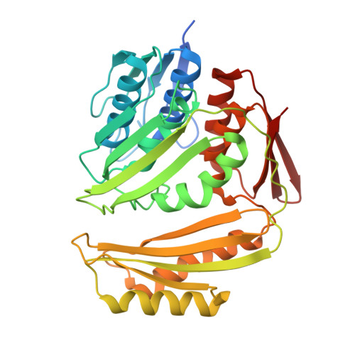
    RNA 3'-phosphate cyclase (RtcA) synthesizes RNA 2',3' cyclic phosphate ends via three steps: reaction with ATP to form a covalent RtcA-(histidinyl-Nε)-AMP intermediate; transfer of adenylate to an RNA 3'-phosphate to form RNA(3')pp(5')A; and attack of the vicinal O2' on the 3'-phosphorus to form a 2',3' cyclic phosphate and release AMP. Here we report the crystal structures of RtcA•ATP, RtcA•ATP•Mn(2+), and RtcA•ATP•Co(2+) substrate complexes and an RtcA•AMP product complex. Together with the structures of RtcA apoenzyme and the covalent RtcA-AMP intermediate, they illuminate the mechanism of nucleotidyl transfer, especially the stereochemical transitions at the AMP phosphate, the critical role of the metal in orienting the PP(i) leaving group of ATP during step 1, and the protein conformational switches that accompany substrate binding and product release. The octahedral metal complex of RtcA•ATP•Mn(2+) includes nonbridging oxygens from each of the ATP phosphates, two waters, and Glu14 as the sole RtcA component. Whereas the RtcA adenylylation step is metal-catalyzed, the subsequent steps in the cyclization pathway are metal-independent.


  • Organizational Affiliation
    • Molecular Biology Program, Sloan-Kettering Institute, 1275 York Avenue, New York, NY 10065, USA.

Macromolecule Content 

  • Total Structure Weight: 39.08 kDa 
  • Atom Count: 2,687 
  • Modeled Residue Count: 336 
  • Deposited Residue Count: 358 
  • Unique protein chains: 1

Macromolecules

Find similar proteins by:|  3D Structure
Entity ID: 1
MoleculeChains  Sequence LengthOrganismDetailsImage
RNA 3'-terminal phosphate cyclase358Escherichia coli K-12Mutation(s): 2 
Gene Names: b4475JW5688rtcAyhgJyhgK
EC: 6.5.1.4
UniProt
Find proteins for P46849 (Escherichia coli (strain K12))
Explore P46849 
Go to UniProtKB:  P46849
Entity Groups
Sequence Clusters30% Identity50% Identity70% Identity90% Identity95% Identity100% Identity
UniProt GroupP46849
Sequence Annotations
Expand
Reference Sequence

Experimental Data & Validation

Experimental Data

  • Method: X-RAY DIFFRACTION
  • Resolution: 2.10 Å
  • R-Value Free:  0.263 (Depositor), 0.256 (DCC) 
  • R-Value Work:  0.207 (Depositor), 0.201 (DCC) 
  • R-Value Observed: 0.210 (Depositor) 
Space Group: P 21 21 2
Unit Cell:
Length ( Å )Angle ( ˚ )
a = 69.66α = 90
b = 82.95β = 90
c = 51.78γ = 90
Software Package:
Software NamePurpose
CBASSdata collection
PHENIXmodel building
PHENIXrefinement
MOSFLMdata reduction
SCALAdata scaling
PHENIXphasing

Structure Validation

View Full Validation Report


Ligand Structure Quality Assessment 


Entry History 

Deposition Data

Revision History  (Full details and data files)

  • Version 1.0: 2011-12-28
    Type: Initial release
  • Version 1.1: 2012-01-18
    Changes: Database references
  • Version 1.2: 2023-09-13
    Changes: Data collection, Database references, Derived calculations, Refinement description