3TUO | pdb_00003tuo

Crystal structure of N-terminal domain of DNA-binding protein satb1


Experimental Data Snapshot

  • Method: X-RAY DIFFRACTION
  • Resolution: 1.70 Å
  • R-Value Free: 
    0.218 (Depositor), 0.214 (DCC) 
  • R-Value Work: 
    0.158 (Depositor), 0.158 (DCC) 
  • R-Value Observed: 
    0.161 (Depositor) 

wwPDB Validation 3D Report Full Report

Validation slider image for 3TUO

This is version 1.2 of the entry. See complete history

Literature

The structural basis for the oligomerization of the N-terminal domain of SATB1

Wang, Z.Yang, X.Chu, X.Zhang, J.Zhou, H.Shen, Y.Long, J.

(2012) Nucleic Acids Res 40: 4193-4202

  • DOI: https://doi.org/10.1093/nar/gkr1284
  • Primary Citation Related Structures: 
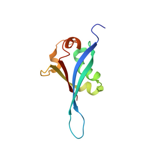
    3TUO

  • PubMed Abstract: 

    Special AT-rich sequence-binding protein 1 (SATB1) is a global chromatin organizer and gene expression regulator essential for T-cell development and breast cancer tumor growth and metastasis. The oligomerization of the N-terminal domain of SATB1 is critical for its biological function. We determined the crystal structure of the N-terminal domain of SATB1. Surprisingly, this domain resembles a ubiquitin domain instead of the previously proposed PDZ domain. Our results also reveal that SATB1 can form a tetramer through its N-terminal domain. The tetramerization of SATB1 plays an essential role in its binding to highly specialized DNA sequences. Furthermore, isothermal titration calorimetry results indicate that the SATB1 tetramer can bind simultaneously to two DNA targets. Based on these results, we propose a molecular model whereby SATB1 regulates the expression of multiple genes both locally and at a distance.


  • Organizational Affiliation
    • State Key Laboratory of Medicinal Chemical Biology and College of Life Sciences, Nankai University, 94 Weijin Road, Tianjin 300071, China.

Macromolecule Content 

  • Total Structure Weight: 46.63 kDa 
  • Atom Count: 3,277 
  • Modeled Residue Count: 391 
  • Deposited Residue Count: 420 
  • Unique protein chains: 1

Macromolecules

Find similar proteins by:|  3D Structure
Entity ID: 1
MoleculeChains  Sequence LengthOrganismDetailsImage
DNA-binding protein SATB1
A, B, C, D
105Homo sapiensMutation(s): 0 
Gene Names: SATB1
UniProt & NIH Common Fund Data Resources
Find proteins for Q01826 (Homo sapiens)
Explore Q01826 
Go to UniProtKB:  Q01826
PHAROS:  Q01826
GTEx:  ENSG00000182568 
Entity Groups
Sequence Clusters30% Identity50% Identity70% Identity90% Identity95% Identity100% Identity
UniProt GroupQ01826
Sequence Annotations
Expand
Reference Sequence

Experimental Data & Validation

Experimental Data

  • Method: X-RAY DIFFRACTION
  • Resolution: 1.70 Å
  • R-Value Free:  0.218 (Depositor), 0.214 (DCC) 
  • R-Value Work:  0.158 (Depositor), 0.158 (DCC) 
  • R-Value Observed: 0.161 (Depositor) 
Space Group: P 21 21 21
Unit Cell:
Length ( Å )Angle ( ˚ )
a = 35.895α = 90
b = 71.027β = 90
c = 153.763γ = 90
Software Package:
Software NamePurpose
MAR345dtbdata collection
PHENIXmodel building
PHENIXrefinement
HKL-2000data reduction
HKL-2000data scaling
PHENIXphasing

Structure Validation

View Full Validation Report


Entry History 

Deposition Data

Revision History  (Full details and data files)

  • Version 1.0: 2012-02-01
    Type: Initial release
  • Version 1.1: 2013-06-26
    Changes: Database references
  • Version 1.2: 2024-03-20
    Changes: Data collection, Database references