3TUC

Crystal structure of SYK kinase domain with 1-benzyl-N-(5-(6,7-dimethoxyquinolin-4-yloxy)pyridin-2-yl)-2-oxo-1,2-dihydropyridine-3-carboxamide


Experimental Data Snapshot

  • Method: X-RAY DIFFRACTION
  • Resolution: 2.10 Å
  • R-Value Free: 0.285 
  • R-Value Work: 0.213 
  • R-Value Observed: 0.216 

wwPDB Validation   3D Report Full Report


Ligand Structure Quality Assessment 


This is version 1.2 of the entry. See complete history


Literature

Identification of Type-II Inhibitors Using Kinase Structures.

Lovering, F.McDonald, J.Whitlock, G.A.Glossop, P.A.Phillips, C.Bent, A.Sabnis, Y.Ryan, M.Fitz, L.Lee, J.Chang, J.S.Han, S.Kurumbail, R.Thorarensen, A.

(2012) Chem Biol Drug Des 80: 657-664

  • DOI: https://doi.org/10.1111/j.1747-0285.2012.01443.x
  • Primary Citation of Related Structures:  
    3TUB, 3TUC, 3TUD

  • PubMed Abstract: 

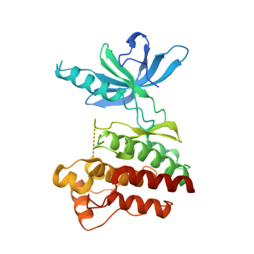
    Spleen tyrosine kinase is a non-receptor tyrosine kinase, overactivation of which is thought to contribute to autoimmune diseases as well as allergy and asthma. Protein kinases have a highly conserved ATP binding site, thus making challenging the design of selective small molecule inhibitors. It has been well documented that some protein kinases can be stabilized in their inactive conformations (Type-II inhibitors). Herein, we describe a protein structure/ligand-based approach to successfully identify ligands that bind to novel conformations of spleen tyrosine kinase. By utilizing kinase protein crystal structures both in the public domain (RCSB) and within Pfizer's protein crystal database, we report the discovery of the first spleen tyrosine kinase Type-II ligands. Compounds 1 and 3 were found to bind to the DFG-out conformation of spleen tyrosine kinase, while compound 2 binds to a DFG-in, C-Helix-out conformation. In this instance, the C-helix moved significantly to create a large hydrophobic pocket rarely seen in kinase protein crystal structures.


  • Organizational Affiliation

    World Wide Medicinal Chemistry, Pfizer Worldwide R & D, 200 Cambridgepark Drive, Cambridge, MA 02140, USA. frank.lovering@pfizer.com


Macromolecules
Find similar proteins by:  (by identity cutoff)  |  3D Structure
Entity ID: 1
MoleculeChains Sequence LengthOrganismDetailsImage
Tyrosine-protein kinase SYK299Homo sapiensMutation(s): 0 
Gene Names: SYK
EC: 2.7.10.2
UniProt & NIH Common Fund Data Resources
Find proteins for P43405 (Homo sapiens)
Explore P43405 
Go to UniProtKB:  P43405
PHAROS:  P43405
GTEx:  ENSG00000165025 
Entity Groups  
Sequence Clusters30% Identity50% Identity70% Identity90% Identity95% Identity100% Identity
UniProt GroupP43405
Sequence Annotations
Expand
  • Reference Sequence
Small Molecules
Ligands 1 Unique
IDChains Name / Formula / InChI Key2D Diagram3D Interactions
FPW
Query on FPW

Download Ideal Coordinates CCD File 
B [auth A]1-benzyl-N-{5-[(6,7-dimethoxyquinolin-4-yl)oxy]pyridin-2-yl}-2-oxo-1,2-dihydropyridine-3-carboxamide
C29 H24 N4 O5
MIRAJAFOEINWJO-UHFFFAOYSA-N
Binding Affinity Annotations 
IDSourceBinding Affinity
FPW Binding MOAD:  3TUC IC50: 100 (nM) from 1 assay(s)
BindingDB:  3TUC IC50: 100 (nM) from 1 assay(s)
Experimental Data & Validation

Experimental Data

  • Method: X-RAY DIFFRACTION
  • Resolution: 2.10 Å
  • R-Value Free: 0.285 
  • R-Value Work: 0.213 
  • R-Value Observed: 0.216 
  • Space Group: P 21 21 21
Unit Cell:
Length ( Å )Angle ( ˚ )
a = 48α = 90
b = 69.47β = 90
c = 83.61γ = 90
Software Package:
Software NamePurpose
HKL-2000data collection
PHASERphasing
BUSTERrefinement
HKL-2000data reduction
SCALAdata scaling

Structure Validation

View Full Validation Report


Ligand Structure Quality Assessment 


Entry History 

Revision History  (Full details and data files)

  • Version 1.0: 2012-08-29
    Type: Initial release
  • Version 1.1: 2012-10-17
    Changes: Database references
  • Version 1.2: 2024-02-28
    Changes: Data collection, Database references, Derived calculations