3TTZ | pdb_00003ttz

Crystal structure of a topoisomerase ATPase inhibitor


Experimental Data Snapshot

  • Method: X-RAY DIFFRACTION
  • Resolution: 1.63 Å
  • R-Value Free: 
    0.194 (Depositor), 0.204 (DCC) 
  • R-Value Work: 
    0.172 (Depositor), 0.186 (DCC) 
  • R-Value Observed: 
    0.174 (Depositor) 

wwPDB Validation 3D Report Full Report

Validation slider image for 3TTZ

Ligand Structure Quality Assessment 


This is version 1.3 of the entry. See complete history

Literature

Pyrrolamide DNA gyrase inhibitors: Optimization of antibacterial activity and efficacy.

Sherer, B.A.Hull, K.Green, O.Basarab, G.Hauck, S.Hill, P.Loch, J.T.Mullen, G.Bist, S.Bryant, J.Boriack-Sjodin, A.Read, J.Degrace, N.Uria-Nickelsen, M.Illingworth, R.N.Eakin, A.E.

(2011) Bioorg Med Chem Lett 21: 7416-7420

  • DOI: https://doi.org/10.1016/j.bmcl.2011.10.010
  • Primary Citation Related Structures: 
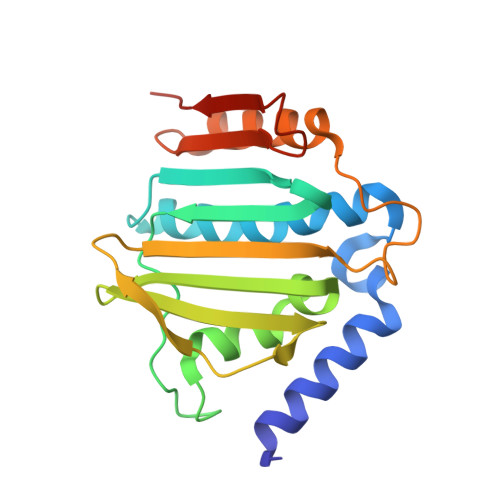
    3TTZ

  • PubMed Abstract: 

    The pyrrolamides are a new class of antibacterial agents targeting DNA gyrase, an essential enzyme across bacterial species and inhibition results in the disruption of DNA synthesis and subsequently, cell death. The optimization of biochemical activity and other drug-like properties through substitutions to the pyrrole, piperidine, and heterocycle portions of the molecule resulted in pyrrolamides with improved cellular activity and in vivo efficacy.


  • Organizational Affiliation
    • Infection Innovative Medicines Unit, AstraZeneca R&D Boston, 35 Gatehouse Drive, Waltham, MA 02451, USA. brian.sherer@astrazeneca.com

Macromolecule Content 

  • Total Structure Weight: 46.05 kDa 
  • Atom Count: 3,562 
  • Modeled Residue Count: 379 
  • Deposited Residue Count: 396 
  • Unique protein chains: 1

Macromolecules

Find similar proteins by:|  3D Structure
Entity ID: 1
MoleculeChains  Sequence LengthOrganismDetailsImage
DNA gyrase subunit B
A, B
198Staphylococcus aureusMutation(s): 0 
Gene Names: gyrB
EC: 5.99.1.3 (PDB Primary Data), 5.6.2.2 (UniProt)
UniProt
Find proteins for P0A0K8 (Staphylococcus aureus)
Explore P0A0K8 
Go to UniProtKB:  P0A0K8
Entity Groups
Sequence Clusters30% Identity50% Identity70% Identity90% Identity95% Identity100% Identity
UniProt GroupP0A0K8
Sequence Annotations
Expand
Reference Sequence

Small Molecules

Ligands 2 Unique
IDChains Name / Formula / InChI Key2D Diagram3D Interactions
07N

Query on 07N


Download:Ideal Coordinates CCD File
C [auth A],
F [auth B]
2-[(3S,4R)-4-{[(3,4-dichloro-5-methyl-1H-pyrrol-2-yl)carbonyl]amino}-3-fluoropiperidin-1-yl]-1,3-thiazole-5-carboxylic acid
C15 H15 Cl2 F N4 O3 S
BGDGHNSKANYMHF-JGVFFNPUSA-N
MG

Query on MG


Download:Ideal Coordinates CCD File
D [auth A],
E [auth A],
G [auth B],
H [auth B],
I [auth B]
MAGNESIUM ION
Mg
JLVVSXFLKOJNIY-UHFFFAOYSA-N
Binding Affinity Annotations 
IDSourceBinding Affinity
07N BindingDB:  3TTZ IC50: min: 4, max: 10 (nM) from 2 assay(s)

Experimental Data & Validation

Experimental Data

  • Method: X-RAY DIFFRACTION
  • Resolution: 1.63 Å
  • R-Value Free:  0.194 (Depositor), 0.204 (DCC) 
  • R-Value Work:  0.172 (Depositor), 0.186 (DCC) 
  • R-Value Observed: 0.174 (Depositor) 
Space Group: C 1 2 1
Unit Cell:
Length ( Å )Angle ( ˚ )
a = 143.178α = 90
b = 55.629β = 100.37
c = 51.216γ = 90
Software Package:
Software NamePurpose
SCALAdata scaling
AMoREphasing
REFMACrefinement
PDB_EXTRACTdata extraction
JDirectordata collection
XSCALEdata scaling

Structure Validation

View Full Validation Report


Ligand Structure Quality Assessment 


Entry History 

Deposition Data

Revision History  (Full details and data files)

  • Version 1.0: 2011-11-16
    Type: Initial release
  • Version 1.1: 2011-12-07
    Changes: Database references
  • Version 1.2: 2017-08-09
    Changes: Refinement description, Source and taxonomy
  • Version 1.3: 2024-02-28
    Changes: Data collection, Database references, Derived calculations