3TRQ

Crystal structure of native rabbit skeletal calsequestrin


Experimental Data Snapshot

  • Method: X-RAY DIFFRACTION
  • Resolution: 1.76 Å
  • R-Value Free: 0.213 
  • R-Value Work: 0.178 
  • R-Value Observed: 0.180 

wwPDB Validation   3D Report Full Report


This is version 2.1 of the entry. See complete history


Literature

Glycosylation of Skeletal Calsequestrin: IMPLICATIONS FOR ITS FUNCTION.

Sanchez, E.J.Lewis, K.M.Munske, G.R.Nissen, M.S.Kang, C.

(2012) J Biol Chem 287: 3042-3050

  • DOI: https://doi.org/10.1074/jbc.M111.326363
  • Primary Citation of Related Structures:  
    3TRQ, 3US3

  • PubMed Abstract: 

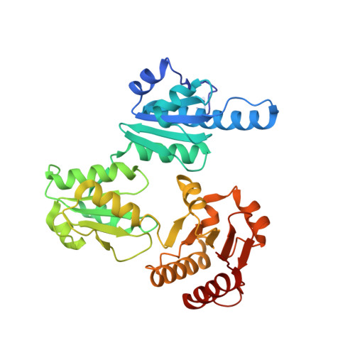
    Calsequestrin (CASQ) serves as a major Ca(2+) storage/buffer protein in the sarcoplasmic reticulum (SR). When purified from skeletal muscle, CASQ1 is obtained in its glycosylated form. Here, we have confirmed the specific site and degree of glycosylation of native rabbit CASQ1 and have investigated its effect on critical properties of CASQ by comparison with the non-glycosylated recombinant form. Based on our comparative approach utilizing crystal structures, Ca(2+) binding capacities, analytical ultracentrifugation, and light-scattering profiles of the native and recombinant rabbit CASQ1, we propose a novel and dynamic role for glycosylation in CASQ. CASQ undergoes a unique degree of mannose trimming as it is trafficked from the proximal endoplasmic reticulum to the SR. The major glycoform of CASQ (GlcNAc(2)Man(9)) found in the proximal endoplasmic reticulum can severely hinder formation of the back-to-back interface, potentially preventing premature Ca(2+)-dependent polymerization of CASQ and ensuring its continuous mobility to the SR. Only trimmed glycans can stabilize both front-to-front and the back-to-back interfaces of CASQ through extensive hydrogen bonding and electrostatic interactions. Therefore, the mature glycoform of CASQ (GlcNAc(2)Man(1-4)) within the SR can be retained upon establishing a functional high capacity Ca(2+) binding polymer. In addition, based on the high resolution structures, we propose a molecular mechanism for the catecholaminergic polymorphic ventricular tachycardia (CPVT2) mutation, K206N.


  • Organizational Affiliation

    School of Molecular Biosciences, Washington State University, Pullman, Washington 99164-4660, USA.


Macromolecules
Find similar proteins by:  (by identity cutoff)  |  3D Structure
Entity ID: 1
MoleculeChains Sequence LengthOrganismDetailsImage
Calsequestrin-1353Oryctolagus cuniculusMutation(s): 0 
UniProt
Find proteins for P07221 (Oryctolagus cuniculus)
Explore P07221 
Go to UniProtKB:  P07221
Entity Groups  
Sequence Clusters30% Identity50% Identity70% Identity90% Identity95% Identity100% Identity
UniProt GroupP07221
Sequence Annotations
Expand
  • Reference Sequence
Oligosaccharides

Help

Entity ID: 2
MoleculeChains Length2D Diagram Glycosylation3D Interactions
alpha-D-mannopyranose-(1-3)-beta-D-mannopyranose-(1-4)-2-acetamido-2-deoxy-beta-D-glucopyranose-(1-4)-2-acetamido-2-deoxy-beta-D-glucopyranose
B
4N-Glycosylation
Glycosylation Resources
GlyTouCan:  G81315DD
GlyCosmos:  G81315DD
GlyGen:  G81315DD
Small Molecules
Ligands 4 Unique
IDChains Name / Formula / InChI Key2D Diagram3D Interactions
MPD
Query on MPD

Download Ideal Coordinates CCD File 
D [auth A],
F [auth A]
(4S)-2-METHYL-2,4-PENTANEDIOL
C6 H14 O2
SVTBMSDMJJWYQN-YFKPBYRVSA-N
MRD
Query on MRD

Download Ideal Coordinates CCD File 
C [auth A],
E [auth A]
(4R)-2-METHYLPENTANE-2,4-DIOL
C6 H14 O2
SVTBMSDMJJWYQN-RXMQYKEDSA-N
CA
Query on CA

Download Ideal Coordinates CCD File 
G [auth A]
H [auth A]
I [auth A]
J [auth A]
K [auth A]
G [auth A],
H [auth A],
I [auth A],
J [auth A],
K [auth A],
L [auth A]
CALCIUM ION
Ca
BHPQYMZQTOCNFJ-UHFFFAOYSA-N
NA
Query on NA

Download Ideal Coordinates CCD File 
M [auth A],
N [auth A],
O [auth A]
SODIUM ION
Na
FKNQFGJONOIPTF-UHFFFAOYSA-N
Experimental Data & Validation

Experimental Data

  • Method: X-RAY DIFFRACTION
  • Resolution: 1.76 Å
  • R-Value Free: 0.213 
  • R-Value Work: 0.178 
  • R-Value Observed: 0.180 
  • Space Group: C 2 2 21
Unit Cell:
Length ( Å )Angle ( ˚ )
a = 59.271α = 90
b = 144.565β = 90
c = 111.17γ = 90
Software Package:
Software NamePurpose
PHENIXrefinement

Structure Validation

View Full Validation Report


Entry History 

Deposition Data

Revision History  (Full details and data files)

  • Version 1.0: 2011-12-21
    Type: Initial release
  • Version 1.1: 2012-01-25
    Changes: Database references
  • Version 1.2: 2012-02-15
    Changes: Database references
  • Version 2.0: 2020-07-29
    Type: Remediation
    Reason: Carbohydrate remediation
    Changes: Advisory, Atomic model, Data collection, Derived calculations, Structure summary
  • Version 2.1: 2023-09-13
    Changes: Data collection, Database references, Refinement description, Structure summary