3TNX | pdb_00003tnx

Structure of the precursor of a thermostable variant of papain at 2.6 Angstroem resolution


Experimental Data Snapshot

  • Method: X-RAY DIFFRACTION
  • Resolution: 2.62 Å
  • R-Value Free: 
    0.236 (Depositor), 0.253 (DCC) 
  • R-Value Work: 
    0.183 (Depositor), 0.200 (DCC) 
  • R-Value Observed: 
    0.186 (Depositor) 

Starting Model: experimental
View more details

wwPDB Validation 3D Report Full Report

Validation slider image for 3TNX

This is version 1.4 of the entry. See complete history

Literature

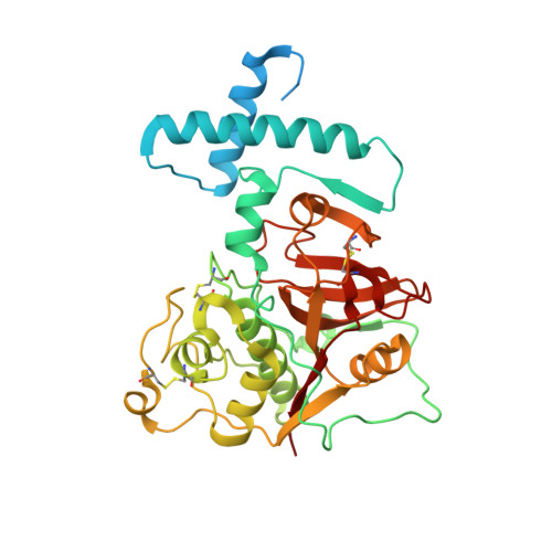
The structure of a thermostable mutant of pro-papain reveals its activation mechanism

Roy, S.Choudhury, D.Aich, P.Dattagupta, J.K.Biswas, S.

(2012) Acta Crystallogr D Biol Crystallogr 68: 1591-1603

  • DOI: https://doi.org/10.1107/S0907444912038607
  • Primary Citation Related Structures: 
    3TNX

  • PubMed Abstract: 

    Papain is the archetype of a broad class of cysteine proteases (clan C1A) that contain a pro-peptide in the zymogen form which is required for correct folding and spatio-temporal regulation of proteolytic activity in the initial stages after expression. This study reports the X-ray structure of the zymogen of a thermostable mutant of papain at 2.6 Å resolution. The overall structure, in particular that of the mature part of the protease, is similar to those of other members of the family. The structure provides an explanation for the molecular basis of the maintenance of latency of the proteolytic activity of the zymogen by its pro-segment at neutral pH. The structural analysis, together with biochemical and biophysical studies, demonstrated that the pro-segment of the zymogen undergoes a rearrangement in the form of a structural loosening at acidic pH which triggers the proteolytic activation cascade. This study further explains the bimolecular stepwise autocatalytic activation mechanism by limited proteolysis of the zymogen of papain at the molecular level. The possible factors responsible for the higher thermal stability of the papain mutant have also been analyzed.


  • Organizational Affiliation
    • Crystallography and Molecular Biology Division, Saha Institute of Nuclear Physics, 1/AF Bidhannagar, Kolkata, India.

Macromolecule Content 

  • Total Structure Weight: 82.17 kDa 
  • Atom Count: 5,079 
  • Modeled Residue Count: 620 
  • Deposited Residue Count: 726 
  • Unique protein chains: 1

Macromolecules

Find similar proteins by:|  3D Structure
Entity ID: 1
MoleculeChains  Sequence LengthOrganismDetailsImage
PapainA,
B [auth C]
363Carica papayaMutation(s): 4 
EC: 3.4.22.2
UniProt
Find proteins for P00784 (Carica papaya)
Explore P00784 
Go to UniProtKB:  P00784
Entity Groups
Sequence Clusters30% Identity50% Identity70% Identity90% Identity95% Identity100% Identity
UniProt GroupP00784
Sequence Annotations
Expand
Reference Sequence

Small Molecules

Ligands 1 Unique
IDChains Name / Formula / InChI Key2D Diagram3D Interactions
CL

Query on CL


Download:Ideal Coordinates CCD File
C [auth A]CHLORIDE ION
Cl
VEXZGXHMUGYJMC-UHFFFAOYSA-M

Experimental Data & Validation

Experimental Data

  • Method: X-RAY DIFFRACTION
  • Resolution: 2.62 Å
  • R-Value Free:  0.236 (Depositor), 0.253 (DCC) 
  • R-Value Work:  0.183 (Depositor), 0.200 (DCC) 
  • R-Value Observed: 0.186 (Depositor) 
Space Group: P 1 21 1
Unit Cell:
Length ( Å )Angle ( ˚ )
a = 42.925α = 90
b = 74.775β = 93.03
c = 116.51γ = 90
Software Package:
Software NamePurpose
PXdata collection
MOLREPphasing
REFMACrefinement
HKL-2000data reduction
SCALEPACKdata scaling

Structure Validation

View Full Validation Report


Entry History 

Deposition Data

Revision History  (Full details and data files)

  • Version 1.0: 2012-09-12
    Type: Initial release
  • Version 1.1: 2012-09-19
    Changes: Database references
  • Version 1.2: 2012-11-28
    Changes: Database references
  • Version 1.3: 2023-11-01
    Changes: Data collection, Database references, Derived calculations, Refinement description
  • Version 1.4: 2024-10-30
    Changes: Structure summary