3TKQ | pdb_00003tkq

Crystal structure of full-length human peroxiredoxin 4 with mixed conformation


Experimental Data Snapshot

  • Method: X-RAY DIFFRACTION
  • Resolution: 2.22 Å
  • R-Value Free: 
    0.224 (Depositor), 0.180 (DCC) 
  • R-Value Work: 
    0.186 (Depositor) 
  • R-Value Observed: 
    0.188 (Depositor) 

Starting Model: experimental
View more details

wwPDB Validation 3D Report Full Report

Validation slider image for 3TKQ

This is version 1.1 of the entry. See complete history

Literature

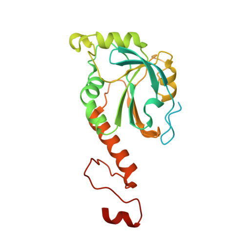
Structural insights into the peroxidase activity and inactivation of human peroxiredoxin 4

Wang, X.Wang, L.Wang, X.Sun, F.Wang, C.-C.

(2011) Biochem J 

  • DOI: https://doi.org/10.1042/BJ20110380
  • Primary Citation Related Structures: 
    3TKP, 3TKQ, 3TKR, 3TKS

  • PubMed Abstract: 

    Prx4 (peroxiredoxin 4) is the only peroxiredoxin located in the ER (endoplasmic reticulum) and a proposed scavenger for H2O2. In the present study, we solved crystal structures of human Prx4 in three different redox forms and characterized the reaction features of Prx4 with H2O2. Prx4 exhibits a toroid-shaped decamer constructed of five catalytic dimers. Structural analysis revealed conformational changes around helix α2 and the C-terminal reigon with a YF (Tyr-Phe) motif from the partner subunit, which are required for interchain disulfide formation between Cys87 and Cys208, a critical step of the catalysis. The structural explanation for the restricting role of the YF motif on the active site dynamics is provided in detail. Prx4 has a high reactivity with H2O2, but is susceptible to overoxidation and consequent inactivation by H2O2. Either deletion of the YF motif or dissociation into dimers decreased the susceptibility of Prx4 to overoxidation by increasing the flexibility of Cys87.


  • Organizational Affiliation
    • National Laboratory of Biomacromolecules, Institute of Biophysics, Chinese Academy of Sciences, Beijing 100101, China.

Macromolecule Content 

  • Total Structure Weight: 140.06 kDa 
  • Atom Count: 7,604 
  • Modeled Residue Count: 873 
  • Deposited Residue Count: 1,230 
  • Unique protein chains: 1

Macromolecules

Find similar proteins by:|  3D Structure
Entity ID: 1
MoleculeChains  Sequence LengthOrganismDetailsImage
Peroxiredoxin-4
A, B, C, D, E
246Homo sapiensMutation(s): 0 
Gene Names: PRDX4
EC: 1.11.1.15 (PDB Primary Data), 1.11.1.24 (UniProt)
UniProt & NIH Common Fund Data Resources
Find proteins for Q13162 (Homo sapiens)
Explore Q13162 
Go to UniProtKB:  Q13162
PHAROS:  Q13162
GTEx:  ENSG00000123131 
Entity Groups
Sequence Clusters30% Identity50% Identity70% Identity90% Identity95% Identity100% Identity
UniProt GroupQ13162
Sequence Annotations
Expand
Reference Sequence

Experimental Data & Validation

Experimental Data

  • Method: X-RAY DIFFRACTION
  • Resolution: 2.22 Å
  • R-Value Free:  0.224 (Depositor), 0.180 (DCC) 
  • R-Value Work:  0.186 (Depositor) 
  • R-Value Observed: 0.188 (Depositor) 
Space Group: C 1 2 1
Unit Cell:
Length ( Å )Angle ( ˚ )
a = 108.486α = 90
b = 139.518β = 103.38
c = 96.197γ = 90
Software Package:
Software NamePurpose
SCALEPACKdata scaling
PHENIXrefinement
PDB_EXTRACTdata extraction
HKL-2000data collection
HKL-2000data reduction
PHASERphasing

Structure Validation

View Full Validation Report


Entry History 

Deposition Data

Revision History  (Full details and data files)

  • Version 1.0: 2011-10-05
    Type: Initial release
  • Version 1.1: 2023-11-01
    Changes: Data collection, Database references, Refinement description