3TGO | pdb_00003tgo

Crystal structure of the E. coli BamCD complex


Experimental Data Snapshot

  • Method: X-RAY DIFFRACTION
  • Resolution: 2.90 Å
  • R-Value Free: 
    0.243 (Depositor), 0.240 (DCC) 
  • R-Value Work: 
    0.177 (Depositor), 0.177 (DCC) 
  • R-Value Observed: 
    0.181 (Depositor) 

Starting Models: experimental
View more details

wwPDB Validation 3D Report Full Report

Validation slider image for 3TGO

This is version 1.2 of the entry. See complete history

Literature

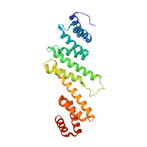
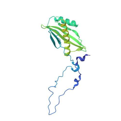
Crystal structure of the beta-barrel assembly machinery BamCD protein complex

Kim, K.H.Aulakh, S.Paetzel, M.

(2011) J Biological Chem 286: 39116-39121

  • DOI: https://doi.org/10.1074/jbc.M111.298166
  • Primary Citation Related Structures: 
    3TGO

  • PubMed Abstract: 

    The β-barrel assembly machinery (BAM) complex of Escherichia coli is a multiprotein machine that catalyzes the essential process of assembling outer membrane proteins. The BAM complex consists of five proteins: one membrane protein, BamA, and four lipoproteins, BamB, BamC, BamD, and BamE. Here, we report the first crystal structure of a Bam lipoprotein complex: the essential lipoprotein BamD in complex with the N-terminal half of BamC (BamC(UN) (Asp(28)-Ala(217)), a 73-residue-long unstructured region followed by the N-terminal domain). The BamCD complex is stabilized predominantly by various hydrogen bonds and salt bridges formed between BamD and the N-terminal unstructured region of BamC. Sequence and molecular surface analyses revealed that many of the conserved residues in both proteins are found at the BamC-BamD interface. A series of truncation mutagenesis and analytical gel filtration chromatography experiments confirmed that the unstructured region of BamC is essential for stabilizing the BamCD complex structure. The unstructured N terminus of BamC interacts with the proposed substrate-binding pocket of BamD, suggesting that this region of BamC may play a regulatory role in outer membrane protein biogenesis.


  • Organizational Affiliation
    • Department of Molecular Biology and Biochemistry, Simon Fraser University, Burnaby, British Columbia V5A 1S6, Canada.

Macromolecule Content 

  • Total Structure Weight: 122.24 kDa 
  • Atom Count: 6,466 
  • Modeled Residue Count: 803 
  • Deposited Residue Count: 1,104 
  • Unique protein chains: 2

Macromolecules

Find similar proteins by:|  3D Structure
Entity ID: 1
MoleculeChains  Sequence LengthOrganismDetailsImage
UPF0169 lipoprotein yfiO
A, B
229Escherichia coli K-12Mutation(s): 0 
Gene Names: b2595JW2577yfiO
Membrane Entity: Yes 
UniProt
Find proteins for P0AC02 (Escherichia coli (strain K12))
Explore P0AC02 
Go to UniProtKB:  P0AC02
Entity Groups
Sequence Clusters30% Identity50% Identity70% Identity90% Identity95% Identity100% Identity
UniProt GroupP0AC02
Sequence Annotations
Expand
Reference Sequence
Find similar proteins by:|  3D Structure
Entity ID: 2
MoleculeChains  Sequence LengthOrganismDetailsImage
Lipoprotein 34
C, D
323Escherichia coli K-12Mutation(s): 0 
Gene Names: b2477dapXJW2462nlpB
Membrane Entity: Yes 
UniProt
Find proteins for P0A903 (Escherichia coli (strain K12))
Explore P0A903 
Go to UniProtKB:  P0A903
Entity Groups
Sequence Clusters30% Identity50% Identity70% Identity90% Identity95% Identity100% Identity
UniProt GroupP0A903
Sequence Annotations
Expand
Reference Sequence

Small Molecules

Ligands 5 Unique
IDChains Name / Formula / InChI Key2D Diagram3D Interactions
PO4

Query on PO4


Download:Ideal Coordinates CCD File
I [auth C]PHOSPHATE ION
O4 P
NBIIXXVUZAFLBC-UHFFFAOYSA-K
GOL

Query on GOL


Download:Ideal Coordinates CCD File
F [auth A],
H [auth B],
K [auth D],
L [auth D]
GLYCEROL
C3 H8 O3
PEDCQBHIVMGVHV-UHFFFAOYSA-N
K

Query on K


Download:Ideal Coordinates CCD File
E [auth A]POTASSIUM ION
K
NPYPAHLBTDXSSS-UHFFFAOYSA-N
CL

Query on CL


Download:Ideal Coordinates CCD File
G [auth B]CHLORIDE ION
Cl
VEXZGXHMUGYJMC-UHFFFAOYSA-M
NA

Query on NA


Download:Ideal Coordinates CCD File
J [auth D]SODIUM ION
Na
FKNQFGJONOIPTF-UHFFFAOYSA-N

Experimental Data & Validation

Experimental Data

  • Method: X-RAY DIFFRACTION
  • Resolution: 2.90 Å
  • R-Value Free:  0.243 (Depositor), 0.240 (DCC) 
  • R-Value Work:  0.177 (Depositor), 0.177 (DCC) 
  • R-Value Observed: 0.181 (Depositor) 
Space Group: I 1 2 1
Unit Cell:
Length ( Å )Angle ( ˚ )
a = 73.78α = 90
b = 133.38β = 100.15
c = 144.992γ = 90
Software Package:
Software NamePurpose
CBASSdata collection
PHASERphasing
REFMACrefinement
MOSFLMdata reduction
SCALAdata scaling

Structure Validation

View Full Validation Report


Entry History 

Deposition Data

Revision History  (Full details and data files)

  • Version 1.0: 2011-09-28
    Type: Initial release
  • Version 1.1: 2011-11-16
    Changes: Database references
  • Version 1.2: 2023-09-13
    Changes: Data collection, Database references, Derived calculations, Refinement description