3TF1

Crystal structure of an H-NOX protein from T. tengcongensis under 6 atm of xenon


Experimental Data Snapshot

  • Method: X-RAY DIFFRACTION
  • Resolution: 2.04 Å
  • R-Value Free: 0.221 
  • R-Value Work: 0.180 
  • R-Value Observed: 0.182 

wwPDB Validation   3D Report Full Report


Ligand Structure Quality Assessment 


This is version 1.1 of the entry. See complete history


Literature

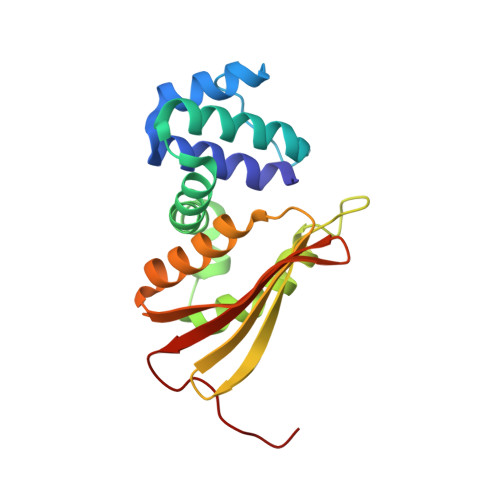
Tunnels modulate ligand flux in a heme nitric oxide/oxygen binding (H-NOX) domain.

Winter, M.B.Herzik, M.A.Kuriyan, J.Marletta, M.A.

(2011) Proc Natl Acad Sci U S A 108: E881-E889

  • DOI: https://doi.org/10.1073/pnas.1114038108
  • Primary Citation of Related Structures:  
    3TF0, 3TF1, 3TF8, 3TF9, 3TFA, 3TFD, 3TFE, 3TFF, 3TFG

  • PubMed Abstract: 

    Interior topological features, such as pockets and channels, have evolved in proteins to regulate biological functions by facilitating the diffusion of biomolecules. Decades of research using the globins as model heme proteins have clearly highlighted the importance of gas pockets around the heme in controlling the capture and release of O(2). However, much less is known about how ligand migration contributes to the diverse functions of other heme protein scaffolds. Heme nitric oxide/oxygen binding (H-NOX) domains are a conserved family of gas-sensing heme proteins with a divergent fold that are critical to numerous signaling pathways. Utilizing X-ray crystallography with xenon, a tunnel network has been shown to serve as a molecular pathway for ligand diffusion. Structure-guided mutagenesis results show that the tunnels have unexpected effects on gas-sensing properties in H-NOX domains. The findings provide insights on how the flux of biomolecules through protein scaffolds modulates protein chemistry.


  • Organizational Affiliation

    Department of Chemistry, California Institute for Quantitative Biosciences, University of California, Berkeley, CA 94720, USA.


Macromolecules
Find similar proteins by:  (by identity cutoff)  |  3D Structure
Entity ID: 1
MoleculeChains Sequence LengthOrganismDetailsImage
Methyl-accepting chemotaxis protein
A, B
188Caldanaerobacter subterraneus subsp. tengcongensisMutation(s): 0 
Gene Names: Tar4TTE0680
UniProt
Find proteins for Q8RBX6 (Caldanaerobacter subterraneus subsp. tengcongensis (strain DSM 15242 / JCM 11007 / NBRC 100824 / MB4))
Explore Q8RBX6 
Go to UniProtKB:  Q8RBX6
Entity Groups  
Sequence Clusters30% Identity50% Identity70% Identity90% Identity95% Identity100% Identity
UniProt GroupQ8RBX6
Sequence Annotations
Expand
  • Reference Sequence
Small Molecules
Ligands 4 Unique
IDChains Name / Formula / InChI Key2D Diagram3D Interactions
HEM
Query on HEM

Download Ideal Coordinates CCD File 
C [auth A],
H [auth B]
PROTOPORPHYRIN IX CONTAINING FE
C34 H32 Fe N4 O4
KABFMIBPWCXCRK-RGGAHWMASA-L
XE
Query on XE

Download Ideal Coordinates CCD File 
E [auth A],
F [auth A],
J [auth B],
K [auth B]
XENON
Xe
FHNFHKCVQCLJFQ-UHFFFAOYSA-N
ACT
Query on ACT

Download Ideal Coordinates CCD File 
G [auth A]ACETATE ION
C2 H3 O2
QTBSBXVTEAMEQO-UHFFFAOYSA-M
OXY
Query on OXY

Download Ideal Coordinates CCD File 
D [auth A],
I [auth B]
OXYGEN MOLECULE
O2
MYMOFIZGZYHOMD-UHFFFAOYSA-N
Experimental Data & Validation

Experimental Data

  • Method: X-RAY DIFFRACTION
  • Resolution: 2.04 Å
  • R-Value Free: 0.221 
  • R-Value Work: 0.180 
  • R-Value Observed: 0.182 
  • Space Group: P 21 21 2
Unit Cell:
Length ( Å )Angle ( ˚ )
a = 80.128α = 90
b = 130.214β = 90
c = 42.81γ = 90
Software Package:
Software NamePurpose
Blu-Icedata collection
PHASERphasing
PHENIXrefinement
HKL-2000data reduction
HKL-2000data scaling

Structure Validation

View Full Validation Report


Ligand Structure Quality Assessment 


Entry History 

Deposition Data

Revision History  (Full details and data files)

  • Version 1.0: 2011-11-09
    Type: Initial release
  • Version 1.1: 2023-09-13
    Changes: Data collection, Database references, Derived calculations, Refinement description