


Crystal structure of risS periplasmic domain at low pH dimer with N-termini ordered
Edwards, T.E., Abendroth, J., Labiuk, S.L., Seattle Structural Genomics Center for Infectious Disease (SSGCID)To be published.
Experimental Data Snapshot
Starting Model: experimental
View more details
wwPDB Validation 3D Report Full Report

Entity ID: 1 | |||||
|---|---|---|---|---|---|
| Molecule | Chains | Sequence Length | Organism | Details | Image |
| Sensor histidine kinase RisS | 127 | Burkholderia pseudomallei 1710b | Mutation(s): 0 Gene Names: BURPS1710b_2508, risS EC: 2.7.13.3 | 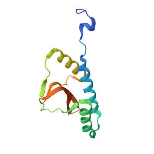 | |
UniProt | |||||
Entity Groups | |||||
| Sequence Clusters | 30% Identity50% Identity70% Identity90% Identity95% Identity100% Identity | ||||
| UniProt Group | Q3JRA3 | ||||
Sequence AnnotationsExpand | |||||
Reference Sequence | |||||
| Length ( Å ) | Angle ( ˚ ) |
|---|---|
| a = 41.06 | α = 90 |
| b = 56.02 | β = 96.03 |
| c = 46.81 | γ = 90 |
| Software Name | Purpose |
|---|---|
| XSCALE | data scaling |
| PHASER | phasing |
| REFMAC | refinement |
| PDB_EXTRACT | data extraction |
| XDS | data reduction |


