3TE7 | pdb_00003te7

Quinone Oxidoreductase (NQ02) bound to the imidazoacridin-6-one 5a1


Experimental Data Snapshot

  • Method: X-RAY DIFFRACTION
  • Resolution: 1.70 Å
  • R-Value Free: 
    0.206 (Depositor), 0.199 (DCC) 
  • R-Value Work: 
    0.170 (Depositor), 0.164 (DCC) 
  • R-Value Observed: 
    0.171 (Depositor) 

Starting Model: experimental
View more details

wwPDB Validation 3D Report Full Report

Validation slider image for 3TE7

Ligand Structure Quality Assessment 


This is version 1.3 of the entry. See complete history

Literature

Novel Inhibitors of NRH:Quinone Oxidoreductase 2 (NQO2): Crystal Structures, Biochemical Activity, and Intracellular Effects of Imidazoacridin-6-ones.

Dunstan, M.S.Barnes, J.Humphries, M.Whitehead, R.C.Bryce, R.A.Leys, D.Stratford, I.J.Nolan, K.A.

(2011) J Med Chem 54: 6597-6611

  • DOI: https://doi.org/10.1021/jm200416e
  • Primary Citation Related Structures: 
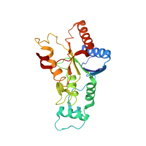
    3TE7, 3TEM

  • PubMed Abstract: 

    Imidazoacridin-6-ones are shown to be potent nanomolar inhibitors of the enzyme NQO2. By use of computational molecular modeling, a reliable QSAR was established, relating inhibitory potency with calculated binding affinity. Further, crystal structures of NQO2 containing two of the imidazoacridin-6-ones have been solved. To generate compounds with reduced off-target (DNA binding) effects, an N-oxide moiety was introduced into the tertiary aminoalkyl side chain of the imidazoacridin-6-ones. This resulted in substantially less toxicity in a panel of eight cancer cell lines, decreased protein binding, and reduced DNA binding and nuclear accumulation. Finally, one of the N-oxides showed potent ability to inhibit the enzymatic function of NQO2 in cells, and therefore, it may be useful as a pharmacological probe to study the properties of the enzyme in vitro and in vivo.


  • Organizational Affiliation
    • Manchester Interdisciplinary Biocentre, University of Manchester and Manchester Cancer Research Centre, Manchester M13 9PT, U.K.

Macromolecule Content 

  • Total Structure Weight: 53.74 kDa 
  • Atom Count: 4,248 
  • Modeled Residue Count: 456 
  • Deposited Residue Count: 456 
  • Unique protein chains: 1

Macromolecules

Find similar proteins by:|  3D Structure
Entity ID: 1
MoleculeChains  Sequence LengthOrganismDetailsImage
Ribosyldihydronicotinamide dehydrogenase [quinone]
A, B
228Homo sapiensMutation(s): 0 
Gene Names: NQO2NMOR2
EC: 1.10.99.2 (PDB Primary Data), 1.10.5.1 (UniProt)
UniProt & NIH Common Fund Data Resources
Find proteins for P16083 (Homo sapiens)
Explore P16083 
Go to UniProtKB:  P16083
PHAROS:  P16083
GTEx:  ENSG00000124588 
Entity Groups
Sequence Clusters30% Identity50% Identity70% Identity90% Identity95% Identity100% Identity
UniProt GroupP16083
Sequence Annotations
Expand
Reference Sequence

Small Molecules

Ligands 4 Unique
IDChains Name / Formula / InChI Key2D Diagram3D Interactions
FAD

Query on FAD


Download:Ideal Coordinates CCD File
D [auth A],
I [auth B]
FLAVIN-ADENINE DINUCLEOTIDE
C27 H33 N9 O15 P2
VWWQXMAJTJZDQX-UYBVJOGSSA-N
TE7

Query on TE7


Download:Ideal Coordinates CCD File
E [auth A],
G [auth B]
5-{[2-(dimethylamino)ethyl]amino}-8-methoxy-6H-imidazo[4,5,1-de]acridin-6-one
C19 H20 N4 O2
NJTQMNXHSDDMHX-UHFFFAOYSA-N
IMD

Query on IMD


Download:Ideal Coordinates CCD File
F [auth A]IMIDAZOLE
C3 H5 N2
RAXXELZNTBOGNW-UHFFFAOYSA-O
ZN

Query on ZN


Download:Ideal Coordinates CCD File
C [auth A],
H [auth B]
ZINC ION
Zn
PTFCDOFLOPIGGS-UHFFFAOYSA-N
Binding Affinity Annotations 
IDSourceBinding Affinity
TE7 BindingDB:  3TE7 IC50: min: 59, max: 3200 (nM) from 2 assay(s)

Experimental Data & Validation

Experimental Data

  • Method: X-RAY DIFFRACTION
  • Resolution: 1.70 Å
  • R-Value Free:  0.206 (Depositor), 0.199 (DCC) 
  • R-Value Work:  0.170 (Depositor), 0.164 (DCC) 
  • R-Value Observed: 0.171 (Depositor) 
Space Group: P 21 21 21
Unit Cell:
Length ( Å )Angle ( ˚ )
a = 56.91α = 90
b = 84.03β = 90
c = 106.55γ = 90
Software Package:
Software NamePurpose
PHENIXrefinement
PDB_EXTRACTdata extraction
ADSCdata collection
XDSdata reduction
XDSdata scaling
PHASERphasing

Structure Validation

View Full Validation Report


Ligand Structure Quality Assessment 


Entry History 

Deposition Data

Revision History  (Full details and data files)

  • Version 1.0: 2011-09-21
    Type: Initial release
  • Version 1.1: 2011-10-05
    Changes: Database references
  • Version 1.2: 2011-10-19
    Changes: Database references
  • Version 1.3: 2023-09-13
    Changes: Data collection, Database references, Derived calculations, Refinement description