3TBD

Crystal Structure of domain VI and LE1 of human Netrin-G2


Experimental Data Snapshot

  • Method: X-RAY DIFFRACTION
  • Resolution: 1.80 Å
  • R-Value Free: 0.227 
  • R-Value Work: 0.175 
  • R-Value Observed: 0.178 

wwPDB Validation   3D Report Full Report


This is version 2.1 of the entry. See complete history


Literature

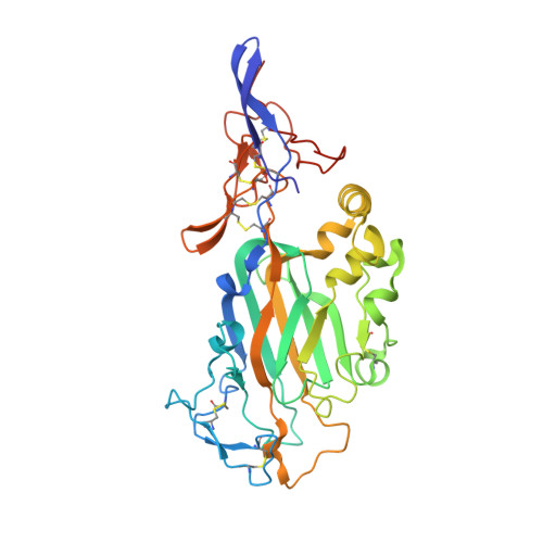
Crystal structure of the ligand binding domain of netrin g2.

Brasch, J.Harrison, O.J.Ahlsen, G.Liu, Q.Shapiro, L.

(2011) J Mol Biol 414: 723-734

  • DOI: https://doi.org/10.1016/j.jmb.2011.10.030
  • Primary Citation of Related Structures:  
    3TBD

  • PubMed Abstract: 

    Netrin G proteins represent a small family of synaptic cell adhesion molecules related to netrins and to the polymerization domains of laminins. Two netrin G proteins are encoded in vertebrate genomes, netrins G1 and G2, which are known to bind the leucine-rich repeat proteins netrin G ligand (NGL)-1 and NGL-2, respectively. Netrin G proteins share a common multi-domain architecture comprising a laminin N-terminal (LN) domain followed by three laminin epidermal growth factor-like (LE) domains and a C' region containing a glycosylphosphatidylinositol anchor. Here, we use deletion analysis to show that the LN domain region of netrin Gs contains the binding site for NGLs to which they bind with 1:1 stoichiometry and sub-micromolar affinity. Netrin Gs are alternatively spliced in their LE domain regions, but the binding region, the LN domain, is identical in all splice forms. We determined the crystal structure for a fragment comprising the LN domain and domain LE1 of netrin G2 by sulfur single-wavelength anomalous diffraction phasing and refined it to 1.8 Å resolution. The structure reveals an overall architecture similar to that of laminin α chain LN domains but includes significant differences including a Ca(2+) binding site in the LN domain. These results reveal the minimal binding unit for interaction of netrin Gs with NGLs, define structural features specific to netrin Gs, and suggest that netrin G alternative splicing is not involved in NGL recognition.


  • Organizational Affiliation

    Department of Biochemistry and Molecular Biophysics, Columbia University, New York, NY 10032, USA.


Macromolecules
Find similar proteins by:  (by identity cutoff)  |  3D Structure
Entity ID: 1
MoleculeChains Sequence LengthOrganismDetailsImage
Netrin-G2338Homo sapiensMutation(s): 1 
Gene Names: NTNG2KIAA1857LMNT2UNQ9381/PRO34206
UniProt & NIH Common Fund Data Resources
Find proteins for Q96CW9 (Homo sapiens)
Explore Q96CW9 
Go to UniProtKB:  Q96CW9
PHAROS:  Q96CW9
GTEx:  ENSG00000196358 
Entity Groups  
Sequence Clusters30% Identity50% Identity70% Identity90% Identity95% Identity100% Identity
UniProt GroupQ96CW9
Sequence Annotations
Expand
  • Reference Sequence
Oligosaccharides

Help

Entity ID: 2
MoleculeChains Length2D Diagram Glycosylation3D Interactions
2-acetamido-2-deoxy-beta-D-glucopyranose-(1-4)-2-acetamido-2-deoxy-beta-D-glucopyranose
B, C
2N-Glycosylation
Glycosylation Resources
GlyTouCan:  G42666HT
GlyCosmos:  G42666HT
GlyGen:  G42666HT
Experimental Data & Validation

Experimental Data

  • Method: X-RAY DIFFRACTION
  • Resolution: 1.80 Å
  • R-Value Free: 0.227 
  • R-Value Work: 0.175 
  • R-Value Observed: 0.178 
  • Space Group: P 32 2 1
Unit Cell:
Length ( Å )Angle ( ˚ )
a = 63.134α = 90
b = 63.134β = 90
c = 190.64γ = 120
Software Package:
Software NamePurpose
HKL-2000data collection
SHELXSphasing
REFMACrefinement
HKL-2000data reduction
SCALEPACKdata scaling

Structure Validation

View Full Validation Report


Entry History 

Deposition Data

Revision History  (Full details and data files)

  • Version 1.0: 2011-11-02
    Type: Initial release
  • Version 1.1: 2012-01-11
    Changes: Database references
  • Version 2.0: 2020-07-29
    Type: Remediation
    Reason: Carbohydrate remediation
    Changes: Atomic model, Data collection, Database references, Derived calculations, Structure summary
  • Version 2.1: 2022-12-21
    Changes: Database references, Structure summary