3T8S | pdb_00003t8s

Apo and InsP3-bound Crystal Structures of the Ligand-Binding Domain of an InsP3 Receptor


Experimental Data Snapshot

  • Method: X-RAY DIFFRACTION
  • Resolution: 3.77 Å
  • R-Value Free: 
    0.292 (Depositor), 0.298 (DCC) 
  • R-Value Work: 
    0.277 (Depositor), 0.273 (DCC) 
  • R-Value Observed: 
    0.278 (Depositor) 

Starting Models: experimental
View more details

wwPDB Validation 3D Report Full Report

Validation slider image for 3T8S

Ligand Structure Quality Assessment 


This is version 1.3 of the entry. See complete history

Literature

Apo and InsP(3)-bound crystal structures of the ligand-binding domain of an InsP(3) receptor.

Lin, C.C.Baek, K.Lu, Z.

(2011) Nat Struct Mol Biol 18: 1172-1174

  • DOI: https://doi.org/10.1038/nsmb.2112
  • Primary Citation Related Structures: 
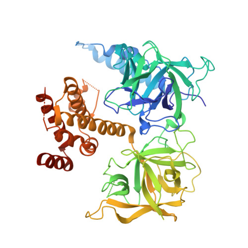
    3T8S

  • PubMed Abstract: 

    We report the crystal structures of the ligand-binding domain (LBD) of a rat inositol 1,4,5-trisphosphate receptor (InsP(3)R) in its apo and InsP(3)-bound conformations. Comparison of these two conformations reveals that LBD's first β-trefoil fold (β-TF1) and armadillo repeat fold (ARF) move together as a unit relative to its second β-trefoil fold (β-TF2). Whereas apo LBD may spontaneously transition between gating conformations, InsP(3) binding shifts this equilibrium toward the active state.


  • Organizational Affiliation
    • Department of Physiology, Howard Hughes Medical Institute, Perelman School of Medicine, University of Pennsylvania, Philadelphia, Pennsylvania, USA.

Macromolecule Content 

  • Total Structure Weight: 132.61 kDa 
  • Atom Count: 7,386 
  • Modeled Residue Count: 983 
  • Deposited Residue Count: 1,170 
  • Unique protein chains: 1

Macromolecules

Find similar proteins by:|  3D Structure
Entity ID: 1
MoleculeChains  Sequence LengthOrganismDetailsImage
Inositol 1,4,5-trisphosphate receptor type 1
A, B
585Rattus norvegicusMutation(s): 0 
Gene Names: Insp3rItpr1
UniProt
Find proteins for P29994 (Rattus norvegicus)
Explore P29994 
Go to UniProtKB:  P29994
Entity Groups
Sequence Clusters30% Identity50% Identity70% Identity90% Identity95% Identity100% Identity
UniProt GroupP29994
Sequence Annotations
Expand
Reference Sequence

Small Molecules

Ligands 1 Unique
IDChains Name / Formula / InChI Key2D Diagram3D Interactions
I3P

Query on I3P


Download:Ideal Coordinates CCD File
C [auth B]D-MYO-INOSITOL-1,4,5-TRIPHOSPHATE
C6 H15 O15 P3
MMWCIQZXVOZEGG-XJTPDSDZSA-N
Binding Affinity Annotations 
IDSourceBinding Affinity
I3P BindingDB:  3T8S Kd: min: 0.21, max: 260 (nM) from 5 assay(s)
IC50: 300 (nM) from 1 assay(s)
EC50: min: 20, max: 209 (nM) from 7 assay(s)

Experimental Data & Validation

Experimental Data

  • Method: X-RAY DIFFRACTION
  • Resolution: 3.77 Å
  • R-Value Free:  0.292 (Depositor), 0.298 (DCC) 
  • R-Value Work:  0.277 (Depositor), 0.273 (DCC) 
  • R-Value Observed: 0.278 (Depositor) 
Space Group: C 1 2 1
Unit Cell:
Length ( Å )Angle ( ˚ )
a = 159.829α = 90
b = 84.877β = 116.92
c = 95.09γ = 90
Software Package:
Software NamePurpose
DENZOdata reduction
SCALEPACKdata scaling
PHASERphasing
REFMACrefinement
PDB_EXTRACTdata extraction
HKL-2000data collection

Structure Validation

View Full Validation Report


Ligand Structure Quality Assessment 


Entry History 

Deposition Data

Revision History  (Full details and data files)

  • Version 1.0: 2011-09-07
    Type: Initial release
  • Version 1.1: 2011-09-21
    Changes: Database references
  • Version 1.2: 2011-10-26
    Changes: Database references
  • Version 1.3: 2023-09-13
    Changes: Data collection, Database references, Derived calculations, Refinement description