3T7L | pdb_00003t7l

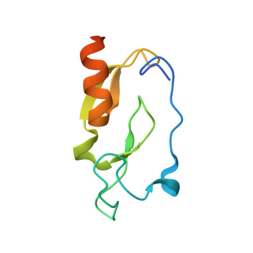
Crystal structure of the FYVE domain of endofin (ZFYVE16) at 1.1A resolution


Experimental Data Snapshot

  • Method: X-RAY DIFFRACTION
  • Resolution: 1.09 Å
  • R-Value Free: 
    0.154 (Depositor), 0.157 (DCC) 
  • R-Value Work: 
    0.142 (Depositor), 0.150 (DCC) 
  • R-Value Observed: 
    0.142 (Depositor) 

wwPDB Validation 3D Report Full Report

Validation slider image for 3T7L

This is version 1.1 of the entry. See complete history

Literature

Macromolecule Content 

  • Total Structure Weight: 10.44 kDa 
  • Atom Count: 745 
  • Modeled Residue Count: 74 
  • Deposited Residue Count: 90 
  • Unique protein chains: 1

Macromolecules

Find similar proteins by:|  3D Structure
Entity ID: 1
MoleculeChains  Sequence LengthOrganismDetailsImage
Zinc finger FYVE domain-containing protein 1690Homo sapiensMutation(s): 0 
Gene Names: KIAA0305ZFYVE16
UniProt & NIH Common Fund Data Resources
Find proteins for Q7Z3T8 (Homo sapiens)
Explore Q7Z3T8 
Go to UniProtKB:  Q7Z3T8
PHAROS:  Q7Z3T8
GTEx:  ENSG00000039319 
Entity Groups
Sequence Clusters30% Identity50% Identity70% Identity90% Identity95% Identity100% Identity
UniProt GroupQ7Z3T8
Sequence Annotations
Expand
Reference Sequence

Experimental Data & Validation

Experimental Data

  • Method: X-RAY DIFFRACTION
  • Resolution: 1.09 Å
  • R-Value Free:  0.154 (Depositor), 0.157 (DCC) 
  • R-Value Work:  0.142 (Depositor), 0.150 (DCC) 
  • R-Value Observed: 0.142 (Depositor) 
Space Group: P 43 21 2
Unit Cell:
Length ( Å )Angle ( ˚ )
a = 42.944α = 90
b = 42.944β = 90
c = 76.879γ = 90
Software Package:
Software NamePurpose
GDAdata collection
SHARPphasing
REFMACrefinement
XDSdata reduction
SCALAdata scaling

Structure Validation

View Full Validation Report


Entry History 

Revision History  (Full details and data files)

  • Version 1.0: 2011-08-31
    Type: Initial release
  • Version 1.1: 2024-02-28
    Changes: Data collection, Database references, Derived calculations