3T5W | pdb_00003t5w

2ME modified human SOD1


Experimental Data Snapshot

  • Method: X-RAY DIFFRACTION
  • Resolution: 1.80 Å
  • R-Value Free: 
    0.241 (Depositor), 0.260 (DCC) 
  • R-Value Work: 
    0.200 (Depositor), 0.219 (DCC) 
  • R-Value Observed: 
    0.202 (Depositor) 

Starting Model: experimental
View more details

wwPDB Validation 3D Report Full Report

Validation slider image for 3T5W

This is version 1.3 of the entry. See complete history

Literature

Structural switching of Cu,Zn-superoxide dismutases at loop VI: insights from the crystal structure of 2-mercaptoethanol-modified enzyme

Ihara, K.Fujiwara, N.Yamaguchi, Y.Torigoe, H.Wakatsuki, S.Taniguchi, N.Suzuki, K.

(2012) Biosci Rep 32: 539-548

  • DOI: https://doi.org/10.1042/BSR20120029
  • Primary Citation Related Structures: 
    3T5W

  • PubMed Abstract: 

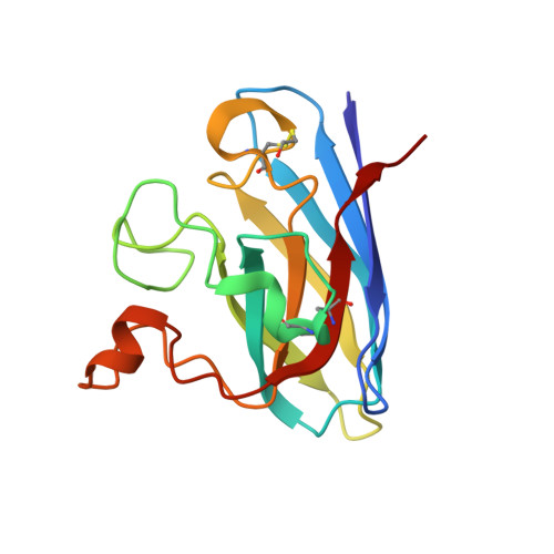
    Cu,Zn SOD1 (superoxide dismutase 1) is implicated in FALS (familial amyotrophic lateral sclerosis) through the accumulation of misfolded proteins that are toxic to neuronal cells. Loop VI (residues 102-115) of the protein is at the dimer interface and could play a critical role in stability. The free cysteine residue, Cys111 in the loop, is readily oxidized and alkylated. We have found that modification of this Cys111 with 2-ME (2-mercaptoethanol; 2-ME-SOD1) stabilizes the protein and the mechanism may provide insights into destabilization and the formation of aggregated proteins. Here, we determined the crystal structure of 2-ME-SOD1 and find that the 2-ME moieties in both subunits interact asymmetrically at the dimer interface and that there is an asymmetric configuration of segment Gly108 to Cys111 in loop VI. One loop VI of the dimer forms a 310-helix (Gly108 to His110) within a unique β-bridge stabilized by a hydrogen bond between Ser105-NH and His110-CO, while the other forms a β-turn without the H-bond. The H-bond (H-type) and H-bond free (F-type) configurations are also seen in some wild-type and mutant human SOD1s in the Protein Data Bank suggesting that they are interconvertible and an intrinsic property of SOD1s. The two structures serve as a basis for classification of these proteins and hopefully a guide to their stability and role in pathophysiology.


  • Organizational Affiliation
    • Structural Biology Research Center, Institute of Material Structure Science, High Energy Accelerator Research Organization, Tsukuba, Ibaraki, Japan.

Macromolecule Content 

  • Total Structure Weight: 193.54 kDa 
  • Atom Count: 15,198 
  • Modeled Residue Count: 1,836 
  • Deposited Residue Count: 1,836 
  • Unique protein chains: 1

Macromolecules

Find similar proteins by:|  3D Structure
Entity ID: 1
MoleculeChains  Sequence LengthOrganismDetailsImage
Superoxide dismutase [Cu-Zn]153Homo sapiensMutation(s): 0 
Gene Names: SOD1
EC: 1.15.1.1 (PDB Primary Data), 1.8 (UniProt)
UniProt & NIH Common Fund Data Resources
Find proteins for P00441 (Homo sapiens)
Explore P00441 
Go to UniProtKB:  P00441
PHAROS:  P00441
GTEx:  ENSG00000142168 
Entity Groups
Sequence Clusters30% Identity50% Identity70% Identity90% Identity95% Identity100% Identity
UniProt GroupP00441
Sequence Annotations
Expand
Reference Sequence

Small Molecules

Ligands 3 Unique
IDChains Name / Formula / InChI Key2D Diagram3D Interactions
SO4

Query on SO4


Download:Ideal Coordinates CCD File
AA [auth F]
DA [auth G]
GA [auth H]
JA [auth I]
MA [auth J]
AA [auth F],
DA [auth G],
GA [auth H],
JA [auth I],
MA [auth J],
O [auth A],
PA [auth K],
R [auth B],
SA [auth L],
U [auth D],
VA [auth M],
X [auth E]
SULFATE ION
O4 S
QAOWNCQODCNURD-UHFFFAOYSA-L
ZN

Query on ZN


Download:Ideal Coordinates CCD File
CA [auth G]
FA [auth H]
IA [auth I]
LA [auth J]
N [auth A]
CA [auth G],
FA [auth H],
IA [auth I],
LA [auth J],
N [auth A],
OA [auth K],
Q [auth B],
RA [auth L],
T [auth D],
UA [auth M],
W [auth E],
Z [auth F]
ZINC ION
Zn
PTFCDOFLOPIGGS-UHFFFAOYSA-N
CU1

Query on CU1


Download:Ideal Coordinates CCD File
BA [auth G]
EA [auth H]
HA [auth I]
KA [auth J]
M [auth A]
BA [auth G],
EA [auth H],
HA [auth I],
KA [auth J],
M [auth A],
NA [auth K],
P [auth B],
QA [auth L],
S [auth D],
TA [auth M],
V [auth E],
Y [auth F]
COPPER (I) ION
Cu
VMQMZMRVKUZKQL-UHFFFAOYSA-N
Modified Residues  1 Unique
IDChains TypeFormula2D DiagramParent
CME
Query on CME
A
B
C [auth D]
D [auth E]
E [auth F]
A,
B,
C [auth D],
D [auth E],
E [auth F],
F [auth G],
G [auth H],
H [auth I],
I [auth J],
J [auth K],
K [auth L],
L [auth M]
L-PEPTIDE LINKINGC5 H11 N O3 S2CYS

Experimental Data & Validation

Experimental Data

  • Method: X-RAY DIFFRACTION
  • Resolution: 1.80 Å
  • R-Value Free:  0.241 (Depositor), 0.260 (DCC) 
  • R-Value Work:  0.200 (Depositor), 0.219 (DCC) 
  • R-Value Observed: 0.202 (Depositor) 
Space Group: P 21 21 21
Unit Cell:
Length ( Å )Angle ( ˚ )
a = 74.086α = 90
b = 163.277β = 90
c = 173.628γ = 90
Software Package:
Software NamePurpose
HKL-2000data collection
MOLREPphasing
REFMACrefinement
HKL-2000data reduction
HKL-2000data scaling

Structure Validation

View Full Validation Report


Entry History 

Deposition Data

Revision History  (Full details and data files)

  • Version 1.0: 2012-08-01
    Type: Initial release
  • Version 1.1: 2013-07-03
    Changes: Database references
  • Version 1.2: 2023-11-01
    Changes: Data collection, Database references, Derived calculations, Refinement description
  • Version 1.3: 2024-10-09
    Changes: Structure summary