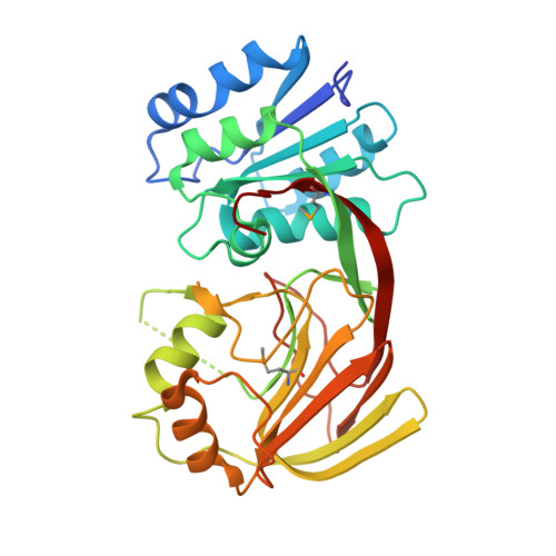
Crystal structure of a putative diacylglycerol kinase from Bacillus anthracis str. Sterne
Hou, J., Zheng, H., Chruszcz, M., Cooper, D.R., Onopriyenko, O., Grimshaw, S., Savchenko, A., Anderson, W.F., Minor, W., Center for Structural Genomics of Infectious Diseases (CSGID)To be published.