3T4O | pdb_00003t4o

Arabidopsis histidine kinase 4 sensor domain in complex with dihydrozeatin


Experimental Data Snapshot

  • Method: X-RAY DIFFRACTION
  • Resolution: 1.75 Å
  • R-Value Free: 
    0.202 (Depositor), 0.209 (DCC) 
  • R-Value Work: 
    0.179 (Depositor), 0.183 (DCC) 
  • R-Value Observed: 
    0.181 (Depositor) 

Starting Model: experimental
View more details

wwPDB Validation 3D Report Full Report

Validation slider image for 3T4O

Ligand Structure Quality Assessment 


This is version 1.3 of the entry. See complete history

Literature

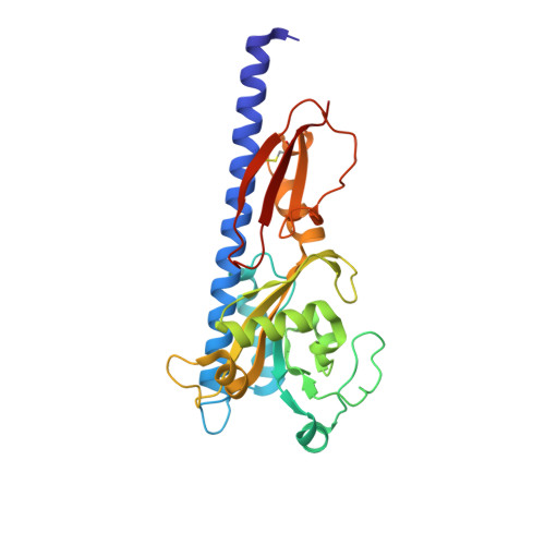
Structural basis for cytokinin recognition by Arabidopsis thaliana histidine kinase 4.

Hothorn, M.Dabi, T.Chory, J.

(2011) Nat Chem Biol 7: 766-768

  • DOI: https://doi.org/10.1038/nchembio.667
  • Primary Citation Related Structures: 
    3T4J, 3T4K, 3T4L, 3T4O, 3T4Q, 3T4S, 3T4T

  • PubMed Abstract: 

    Cytokinins are classic hormones that orchestrate plant growth and development and the integrity of stem cell populations. Cytokinin receptors are eukaryotic sensor histidine kinases that are activated by both naturally occurring adenine-type cytokinins and urea-based synthetic compounds. Crystal structures of the Arabidopsis thaliana histidine kinase 4 sensor domain in complex with different cytokinin ligands now rationalize the hormone-binding specificity of the receptor and may spur the design of new cytokinin ligands.


  • Organizational Affiliation
    • Plant Biology Laboratory, The Salk Institute for Biological Studies, La Jolla, California, USA.

Macromolecule Content 

  • Total Structure Weight: 62.02 kDa 
  • Atom Count: 4,991 
  • Modeled Residue Count: 531 
  • Deposited Residue Count: 540 
  • Unique protein chains: 1

Macromolecules

Find similar proteins by:|  3D Structure
Entity ID: 1
MoleculeChains  Sequence LengthOrganismDetailsImage
Histidine kinase 4
A, B
270Arabidopsis thalianaMutation(s): 0 
Gene Names: AHK4At2g01830CRE1RAW1T23K3.2WOL
EC: 2.7.13.3 (PDB Primary Data), 3.1.3.16 (PDB Primary Data)
UniProt
Find proteins for Q9C5U0 (Arabidopsis thaliana)
Explore Q9C5U0 
Go to UniProtKB:  Q9C5U0
Entity Groups
Sequence Clusters30% Identity50% Identity70% Identity90% Identity95% Identity100% Identity
UniProt GroupQ9C5U0
Sequence Annotations
Expand
Reference Sequence

Experimental Data & Validation

Experimental Data

  • Method: X-RAY DIFFRACTION
  • Resolution: 1.75 Å
  • R-Value Free:  0.202 (Depositor), 0.209 (DCC) 
  • R-Value Work:  0.179 (Depositor), 0.183 (DCC) 
  • R-Value Observed: 0.181 (Depositor) 
Space Group: P 32 2 1
Unit Cell:
Length ( Å )Angle ( ˚ )
a = 60.01α = 90
b = 60.01β = 90
c = 297.86γ = 120
Software Package:
Software NamePurpose
ADSCdata collection
PHASERphasing
REFMACrefinement
XDSdata reduction
XSCALEdata scaling

Structure Validation

View Full Validation Report


Ligand Structure Quality Assessment 


Entry History 

Deposition Data

  • Released Date: 2011-10-05 
  • Deposition Author(s): Hothorn, M.

Revision History  (Full details and data files)

  • Version 1.0: 2011-10-05
    Type: Initial release
  • Version 1.1: 2011-12-14
    Changes: Database references
  • Version 1.2: 2023-09-13
    Changes: Data collection, Database references, Derived calculations, Refinement description
  • Version 1.3: 2024-11-20
    Changes: Structure summary