3T3P | pdb_00003t3p

A Novel High Affinity Integrin alphaIIbbeta3 Receptor Antagonist That Unexpectedly Displaces Mg2+ from the beta3 MIDAS


Experimental Data Snapshot

  • Method: X-RAY DIFFRACTION
  • Resolution: 2.20 Å
  • R-Value Free: 
    0.220 (Depositor), 0.218 (DCC) 
  • R-Value Work: 
    0.189 (Depositor), 0.182 (DCC) 
  • R-Value Observed: 
    0.189 (Depositor) 

wwPDB Validation 3D Report Full Report

Validation slider image for 3T3P

Ligand Structure Quality Assessment 


This is version 2.1 of the entry. See complete history

Literature

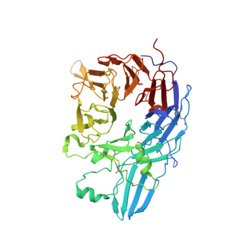
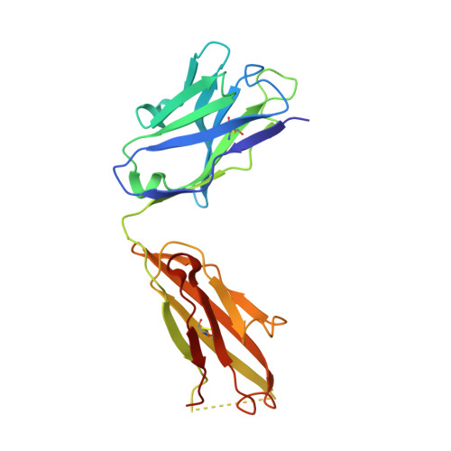
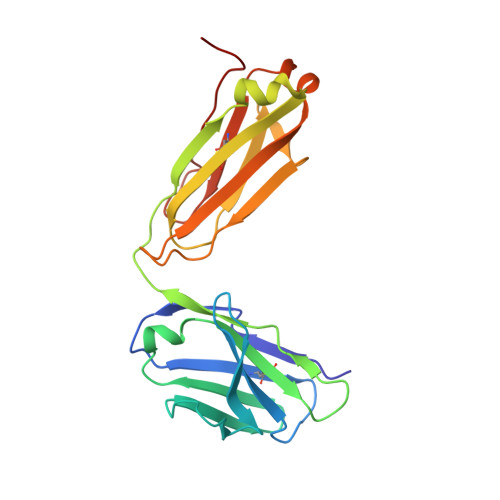
Structure-Guided Design of a High-Affinity Platelet Integrin alphaIIbbeta3 Receptor Antagonist That Disrupts Mg2+ Binding to the MIDAS

Zhu, J.Choi, W.S.McCoy, J.G.Negri, A.Zhu, J.Naini, S.Li, J.Shen, M.Huang, W.Bougie, D.Rasmussen, M.Aster, R.Thomas, C.J.Filizola, M.Springer, T.A.Coller, B.S.

(2012) Sci Transl Med 4: 125ra32-125ra32

  • DOI: https://doi.org/10.1126/scitranslmed.3003576
  • Primary Citation Related Structures: 
    3T3M, 3T3P

  • PubMed Abstract: 

    An integrin found on platelets, α(IIb)β(3) mediates platelet aggregation, and α(IIb)β(3) antagonists are effective antithrombotic agents in the clinic. Ligands bind to integrins in part by coordinating a magnesium ion (Mg(2+)) located in the β subunit metal ion-dependent adhesion site (MIDAS). Drugs patterned on the integrin ligand sequence Arg-Gly-Asp have a basic moiety that binds the α(IIb) subunit and a carboxyl group that coordinates the MIDAS Mg(2+) in the β(3) subunits. They induce conformational changes in the β(3) subunit that may have negative consequences such as exposing previously hidden epitopes and inducing the active conformation of the receptor. We recently reported an inhibitor of α(IIb)β(3) (RUC-1) that binds exclusively to the α(IIb) subunit; here, we report the structure-based design and synthesis of RUC-2, a RUC-1 derivative with a ~100-fold higher affinity. RUC-2 does not induce major conformational changes in β(3) as judged by monoclonal antibody binding, light scattering, gel chromatography, electron microscopy, and a receptor priming assay. X-ray crystallography of the RUC-2-α(IIb)β(3) headpiece complex in 1 mM calcium ion (Ca(2+))/5 mM Mg(2+) at 2.6 Å revealed that RUC-2 binds to α(IIb) the way RUC-1 does, but in addition, it binds to the β(3) MIDAS residue glutamic acid 220, thus displacing Mg(2+) from the MIDAS. When the Mg(2+) concentration was increased to 20 mM, however, Mg(2+) was identified in the MIDAS and RUC-2 was absent. RUC-2's ability to inhibit ligand binding and platelet aggregation was diminished by increasing the Mg(2+) concentration. Thus, RUC-2 inhibits ligand binding by a mechanism different from that of all other α(IIb)β(3) antagonists and may offer advantages as a therapeutic agent.


  • Organizational Affiliation
    • Immune Disease Institute, Children's Hospital Boston, and Department of Pathology, Harvard Medical School, Boston, MA 02115, USA.

Macromolecule Content 

  • Total Structure Weight: 301.79 kDa 
  • Atom Count: 22,411 
  • Modeled Residue Count: 2,702 
  • Deposited Residue Count: 2,728 
  • Unique protein chains: 4

Macromolecules

Find similar proteins by:|  3D Structure
Entity ID: 1
MoleculeChains  Sequence LengthOrganismDetailsImage
Integrin alpha-IIb
A, C
457Homo sapiensMutation(s): 0 
Gene Names: ITGA2BGP2BITGAB
UniProt & NIH Common Fund Data Resources
Find proteins for P08514 (Homo sapiens)
Explore P08514 
Go to UniProtKB:  P08514
PHAROS:  P08514
GTEx:  ENSG00000005961 
Entity Groups
Sequence Clusters30% Identity50% Identity70% Identity90% Identity95% Identity100% Identity
UniProt GroupP08514
Sequence Annotations
Expand
Reference Sequence
Find similar proteins by:|  3D Structure
Entity ID: 2
MoleculeChains  Sequence LengthOrganismDetailsImage
Integrin beta-3
B, D
472Homo sapiensMutation(s): 0 
Gene Names: ITGB3GP3A
UniProt & NIH Common Fund Data Resources
Find proteins for P05106 (Homo sapiens)
Explore P05106 
Go to UniProtKB:  P05106
PHAROS:  P05106
GTEx:  ENSG00000259207 
Entity Groups
Sequence Clusters30% Identity50% Identity70% Identity90% Identity95% Identity100% Identity
UniProt GroupP05106
Glycosylation
Glycosylation Sites: 3Go to GlyGen: P05106-1
Sequence Annotations
Expand
Reference Sequence
Find similar proteins by:|  3D Structure
Entity ID: 3
MoleculeChains  Sequence LengthOrganismDetailsImage
Monoclonal antibody 10E5 heavy chainE,
G [auth H]
221Mus musculusMutation(s): 0 
Find similar proteins by:|  3D Structure
Entity ID: 4
MoleculeChains  Sequence LengthOrganismDetailsImage
Monoclonal antibody 10E5 light chainF,
H [auth L]
214Mus musculusMutation(s): 0 

Oligosaccharides

Help  
Entity ID: 5
MoleculeChains Length2D Diagram GlycosylationD Interactions
alpha-D-mannopyranose-(1-3)-[alpha-D-mannopyranose-(1-6)]beta-D-mannopyranose-(1-4)-2-acetamido-2-deoxy-beta-D-glucopyranose-(1-4)-2-acetamido-2-deoxy-beta-D-glucopyranoseI [auth G]5N-Glycosylation
Glycosylation Resources
GlyTouCan: G22768VO
GlyCosmos: G22768VO
GlyGen: G22768VO
Entity ID: 6
MoleculeChains Length2D Diagram GlycosylationD Interactions
2-acetamido-2-deoxy-beta-D-glucopyranose-(1-4)-2-acetamido-2-deoxy-beta-D-glucopyranoseJ [auth I],
L [auth K]
2N-Glycosylation
Glycosylation Resources
GlyTouCan: G42666HT
GlyCosmos: G42666HT
GlyGen: G42666HT
Entity ID: 7
MoleculeChains Length2D Diagram GlycosylationD Interactions
alpha-D-mannopyranose-(1-3)-beta-D-mannopyranose-(1-4)-2-acetamido-2-deoxy-beta-D-glucopyranose-(1-4)-2-acetamido-2-deoxy-beta-D-glucopyranoseK [auth J]4N-Glycosylation
Glycosylation Resources
GlyTouCan: G81315DD
GlyCosmos: G81315DD
GlyGen: G81315DD

Small Molecules

Ligands 6 Unique
IDChains Name / Formula / InChI Key2D Diagram3D Interactions
NAG

Query on NAG


Download:Ideal Coordinates CCD File
MA [auth D],
Y [auth B]
2-acetamido-2-deoxy-beta-D-glucopyranose
C8 H15 N O6
OVRNDRQMDRJTHS-FMDGEEDCSA-N
SO4

Query on SO4


Download:Ideal Coordinates CCD File
AA [auth C]
BA [auth C]
M [auth A]
N [auth A]
NA [auth L]
AA [auth C],
BA [auth C],
M [auth A],
N [auth A],
NA [auth L],
O [auth A],
Z [auth C]
SULFATE ION
O4 S
QAOWNCQODCNURD-UHFFFAOYSA-L
GOL

Query on GOL


Download:Ideal Coordinates CCD File
P [auth A]GLYCEROL
C3 H8 O3
PEDCQBHIVMGVHV-UHFFFAOYSA-N
CA

Query on CA


Download:Ideal Coordinates CCD File
EA [auth C]
FA [auth C]
GA [auth C]
HA [auth C]
KA [auth D]
EA [auth C],
FA [auth C],
GA [auth C],
HA [auth C],
KA [auth D],
LA [auth D],
Q [auth A],
R [auth A],
S [auth A],
T [auth A],
W [auth B],
X [auth B]
CALCIUM ION
Ca
BHPQYMZQTOCNFJ-UHFFFAOYSA-N
CL

Query on CL


Download:Ideal Coordinates CCD File
CA [auth C],
DA [auth C],
IA [auth D],
U [auth B]
CHLORIDE ION
Cl
VEXZGXHMUGYJMC-UHFFFAOYSA-M
MG

Query on MG


Download:Ideal Coordinates CCD File
JA [auth D],
V [auth B]
MAGNESIUM ION
Mg
JLVVSXFLKOJNIY-UHFFFAOYSA-N

Experimental Data & Validation

Experimental Data

  • Method: X-RAY DIFFRACTION
  • Resolution: 2.20 Å
  • R-Value Free:  0.220 (Depositor), 0.218 (DCC) 
  • R-Value Work:  0.189 (Depositor), 0.182 (DCC) 
  • R-Value Observed: 0.189 (Depositor) 
Space Group: P 21 21 2
Unit Cell:
Length ( Å )Angle ( ˚ )
a = 259.533α = 90
b = 145.257β = 90
c = 104.798γ = 90
Software Package:
Software NamePurpose
Blu-Icedata collection
PHASERphasing
PHENIXrefinement
XDSdata reduction
XDSdata scaling

Structure Validation

View Full Validation Report


Ligand Structure Quality Assessment 


Entry History 

Deposition Data

Revision History  (Full details and data files)

  • Version 1.0: 2012-03-28
    Type: Initial release
  • Version 1.1: 2017-10-25
    Changes: Advisory, Database references, Derived calculations, Refinement description, Source and taxonomy, Structure summary
  • Version 2.0: 2020-07-29
    Type: Remediation
    Reason: Carbohydrate remediation
    Changes: Advisory, Atomic model, Data collection, Derived calculations, Structure summary
  • Version 2.1: 2024-11-27
    Changes: Advisory, Data collection, Database references, Structure summary