3T1I | pdb_00003t1i

Crystal Structure of Human Mre11: Understanding Tumorigenic Mutations


Experimental Data Snapshot

  • Method: X-RAY DIFFRACTION
  • Resolution: 3.00 Å
  • R-Value Free: 
    0.250 (Depositor), 0.248 (DCC) 
  • R-Value Work: 
    0.227 (Depositor), 0.226 (DCC) 
  • R-Value Observed: 
    0.228 (Depositor) 

wwPDB Validation 3D Report Full Report

Validation slider image for 3T1I

This is version 1.1 of the entry. See complete history

Literature

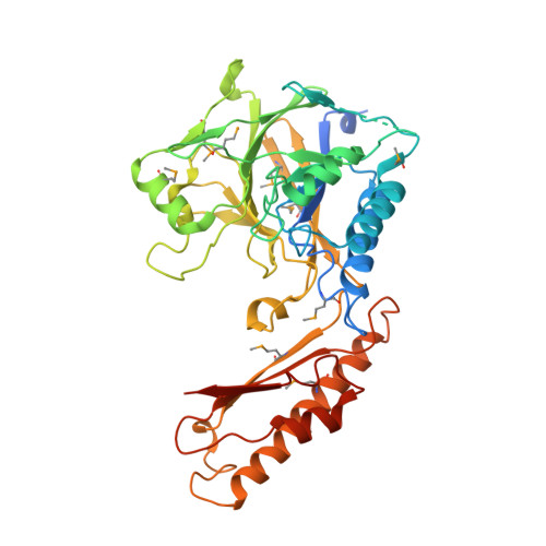
Crystal structure of human mre11: understanding tumorigenic mutations

Park, Y.B.Chae, J.Kim, Y.Cho, Y.

(2011) Structure 19: 1591-1602

  • DOI: https://doi.org/10.1016/j.str.2011.09.010
  • Primary Citation Related Structures: 
    3T1I

  • PubMed Abstract: 

    Mre11 plays an important role in repairing damaged DNA by cleaving broken ends and by providing a platform for other DNA repair proteins. Various Mre11 mutations have been identified in several types of cancer. We have determined the crystal structure of the human Mre11 core (hMre11), which contains the nuclease and capping domains. hMre11 dimerizes through the interfaces between loop β3-α3 from one Mre11 and loop β4-β5 from another Mre11, and between loop α2-β3 from one Mre11 and helices α2 and α3 from another Mre11, and assembles into a completely different dimeric architecture compared with bacterial or archaeal Mre11 homologs. Nbs1 binds to the region containing loop α2-β3 which participates in dimerization. The hMre11 structure in conjunction with biochemical analyses reveals that many tumorigenic mutations are primarily associated with Nbs1 binding and partly with nuclease activities, providing a framework for understanding how mutations inactivate Mre11.


  • Organizational Affiliation
    • Department of Life Science, Pohang University of Science and Technology, Pohang 790-784, South Korea.

Macromolecule Content 

  • Total Structure Weight: 201.43 kDa 
  • Atom Count: 12,407 
  • Modeled Residue Count: 1,517 
  • Deposited Residue Count: 1,724 
  • Unique protein chains: 1

Macromolecules

Find similar proteins by:|  3D Structure
Entity ID: 1
MoleculeChains  Sequence LengthOrganismDetailsImage
Double-strand break repair protein MRE11A
A, B, C, D
431Homo sapiensMutation(s): 0 
Gene Names: HNGS1MRE11MRE11A
EC: 3.1
UniProt & NIH Common Fund Data Resources
Find proteins for P49959 (Homo sapiens)
Explore P49959 
Go to UniProtKB:  P49959
PHAROS:  P49959
GTEx:  ENSG00000020922 
Entity Groups
Sequence Clusters30% Identity50% Identity70% Identity90% Identity95% Identity100% Identity
UniProt GroupP49959
Sequence Annotations
Expand
Reference Sequence

Small Molecules

Ligands 3 Unique
IDChains Name / Formula / InChI Key2D Diagram3D Interactions
DTT

Query on DTT


Download:Ideal Coordinates CCD File
G [auth A],
H [auth A]
2,3-DIHYDROXY-1,4-DITHIOBUTANE
C4 H10 O2 S2
VHJLVAABSRFDPM-IMJSIDKUSA-N
GOL

Query on GOL


Download:Ideal Coordinates CCD File
AA [auth D]
BA [auth D]
CA [auth D]
I [auth A]
J [auth A]
AA [auth D],
BA [auth D],
CA [auth D],
I [auth A],
J [auth A],
K [auth A],
L [auth A],
M [auth A],
N [auth A],
S [auth C],
T [auth C],
U [auth C],
V [auth C],
W [auth C],
X [auth C]
GLYCEROL
C3 H8 O3
PEDCQBHIVMGVHV-UHFFFAOYSA-N
MN

Query on MN


Download:Ideal Coordinates CCD File
E [auth A]
F [auth A]
O [auth B]
P [auth B]
Q [auth C]
E [auth A],
F [auth A],
O [auth B],
P [auth B],
Q [auth C],
R [auth C],
Y [auth D],
Z [auth D]
MANGANESE (II) ION
Mn
WAEMQWOKJMHJLA-UHFFFAOYSA-N
Modified Residues  1 Unique
IDChains TypeFormula2D DiagramParent
MSE
Query on MSE
A, B, C, D
L-PEPTIDE LINKINGC5 H11 N O2 SeMET

Experimental Data & Validation

Experimental Data

  • Method: X-RAY DIFFRACTION
  • Resolution: 3.00 Å
  • R-Value Free:  0.250 (Depositor), 0.248 (DCC) 
  • R-Value Work:  0.227 (Depositor), 0.226 (DCC) 
  • R-Value Observed: 0.228 (Depositor) 
Space Group: P 21 21 21
Unit Cell:
Length ( Å )Angle ( ˚ )
a = 134.79α = 90
b = 135.21β = 90
c = 135.38γ = 90
Software Package:
Software NamePurpose
HKL-2000data collection
CNSrefinement
PHENIXrefinement
HKL-2000data reduction
HKL-2000data scaling
CNSphasing

Structure Validation

View Full Validation Report


Entry History 

Deposition Data

Revision History  (Full details and data files)

  • Version 1.0: 2011-11-30
    Type: Initial release
  • Version 1.1: 2024-11-20
    Changes: Data collection, Database references, Derived calculations, Structure summary