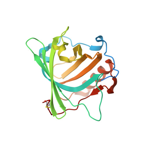
3T1D

The mutant structure of human Siderocalin W79A, R81A, Y106F bound to Enterobactin


Experimental Data Snapshot

  • Method: X-RAY DIFFRACTION
  • Resolution: 2.30 Å
  • R-Value Free: 0.228 
  • R-Value Work: 0.189 
  • R-Value Observed: 0.192 

Starting Model: experimental
View more details

wwPDB Validation   3D Report Full Report


Ligand Structure Quality Assessment 


This is version 1.6 of the entry. See complete history


Literature

Parsing the functional specificity of Siderocalin/Lipocalin 2/NGAL for siderophores and related small-molecule ligands.

Clifton, M.C.Rupert, P.B.Hoette, T.M.Raymond, K.N.Abergel, R.J.Strong, R.K.

(2019) J Struct Biol X 2: 100008-100008

  • DOI: https://doi.org/10.1016/j.yjsbx.2019.100008
  • Primary Citation of Related Structures:  
    3T1D, 6O5D

  • PubMed Abstract: 

    Siderocalin/Lipocalin 2/Neutrophil Gelatinase Associated Lipocalin/24p3 is an innate immune system protein with bacteriostatic activity, acting by tightly binding and sequestering diverse catecholate and mixed-type ferric siderophores from enteric bacteria and mycobacteria. Bacterial virulence achieved through siderophore modifications, or utilization of alternate siderophores, can be explained by evasion of Siderocalin binding. Siderocalin has also been implicated in a wide variety of disease processes, though often in seemingly contradictory ways, and has been proposed to bind to a broader array of ligands beyond siderophores. Using structural, directed mutational, and binding studies, we have sought to rigorously test, and fully elucidate, the Siderocalin recognition mechanism. Several proposed ligands fail to meet rigorous binding criteria, including the bacterial siderophore pyochelin, the iron-chelating catecholamine hormone norepinephrine, and the bacterial second messenger cyclic diguanylate monophosphate. While possessing a remarkably rigid structure, in principle simplifying analyses of ligand recognition, understanding Scn recognition is complicated by the observed conformational and stoichiometric plasticity, and instability, of its bona fide siderophore ligands. Since the role of Siderocalin at the early host/pathogen interface is to compete for bacterial ferric siderophores, we also analyzed how bacterial siderophore binding proteins and enzymes alternately recognize siderophores that efficiently bind to, or evade, Siderocalin sequestration - including determining the crystal structure of Bacillus cereus YfiY bound to schizokinen. These studies combine to refine the potential physiological functions of Siderocalin by defining its multiplexed recognition mechanism.


  • Organizational Affiliation

    Division of Basic Science, Fred Hutchinson Cancer Research Center, Seattle, WA 98109, USA.


Macromolecules
Find similar proteins by:  (by identity cutoff)  |  3D Structure
Entity ID: 1
MoleculeChains Sequence LengthOrganismDetailsImage
Neutrophil gelatinase-associated lipocalin
A, B, C
198Homo sapiensMutation(s): 3 
Gene Names: LCN2HNLNGAL
UniProt & NIH Common Fund Data Resources
Find proteins for P80188 (Homo sapiens)
Explore P80188 
Go to UniProtKB:  P80188
PHAROS:  P80188
GTEx:  ENSG00000148346 
Entity Groups  
Sequence Clusters30% Identity50% Identity70% Identity90% Identity95% Identity100% Identity
UniProt GroupP80188
Sequence Annotations
Expand
  • Reference Sequence
Small Molecules
Ligands 6 Unique
IDChains Name / Formula / InChI Key2D Diagram3D Interactions
TD1
Query on TD1

Download Ideal Coordinates CCD File 
K [auth A]O-[(2S)-2-amino-3-hydroxypropanoyl]-N-(2,3-dihydroxybenzoyl)-L-serine
C13 H16 N2 O8
NNOZTAPXJBUXBD-YUMQZZPRSA-N
DBS
Query on DBS

Download Ideal Coordinates CCD File 
O [auth B],
Q [auth C]
2-(2,3-DIHYDROXY-BENZOYLAMINO)-3-HYDROXY-PROPIONIC ACID
C10 H11 N O6
VDTYHTVHFIIEIL-LURJTMIESA-N
DBH
Query on DBH

Download Ideal Coordinates CCD File 
P [auth B]2,3-DIHYDROXY-BENZOIC ACID
C7 H6 O4
GLDQAMYCGOIJDV-UHFFFAOYSA-N
GOL
Query on GOL

Download Ideal Coordinates CCD File 
D [auth A]
G [auth A]
I [auth A]
J [auth A]
M [auth B]
D [auth A],
G [auth A],
I [auth A],
J [auth A],
M [auth B],
N [auth B],
R [auth C]
GLYCEROL
C3 H8 O3
PEDCQBHIVMGVHV-UHFFFAOYSA-N
CL
Query on CL

Download Ideal Coordinates CCD File 
E [auth A],
F [auth A],
H [auth A]
CHLORIDE ION
Cl
VEXZGXHMUGYJMC-UHFFFAOYSA-M
UNX
Query on UNX

Download Ideal Coordinates CCD File 
L [auth A]UNKNOWN ATOM OR ION
X
Experimental Data & Validation

Experimental Data

  • Method: X-RAY DIFFRACTION
  • Resolution: 2.30 Å
  • R-Value Free: 0.228 
  • R-Value Work: 0.189 
  • R-Value Observed: 0.192 
  • Space Group: P 41 21 2
Unit Cell:
Length ( Å )Angle ( ˚ )
a = 114.749α = 90
b = 114.749β = 90
c = 119.065γ = 90
Software Package:
Software NamePurpose
DENZOdata reduction
SCALEPACKdata scaling
REFMACrefinement
PDB_EXTRACTdata extraction
HKL-2000data reduction
HKL-2000data scaling
REFMACphasing

Structure Validation

View Full Validation Report


Ligand Structure Quality Assessment 


Entry History 

Revision History  (Full details and data files)

  • Version 1.0: 2011-08-17
    Type: Initial release
  • Version 1.1: 2013-10-09
    Changes: Database references
  • Version 1.2: 2017-11-08
    Changes: Refinement description
  • Version 1.3: 2018-01-24
    Changes: Database references
  • Version 1.4: 2019-02-27
    Changes: Data collection, Database references
  • Version 1.5: 2023-07-26
    Changes: Database references, Derived calculations
  • Version 1.6: 2023-09-13
    Changes: Data collection, Refinement description