3T10 | pdb_00003t10

HSP90 N-terminal domain bound to ACP


Experimental Data Snapshot

  • Method: X-RAY DIFFRACTION
  • Resolution: 1.24 Å
  • R-Value Free: 
    0.172 (Depositor), 0.164 (DCC) 
  • R-Value Work: 
    0.158 (Depositor), 0.151 (DCC) 
  • R-Value Observed: 
    0.158 (Depositor) 

Starting Model: experimental
View more details

wwPDB Validation 3D Report Full Report

Validation slider image for 3T10

Ligand Structure Quality Assessment 


This is version 1.2 of the entry. See complete history

Literature

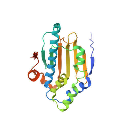
Structure insights into mechanisms of ATP hydrolysis and the activation of human heat-shock protein 90.

Li, J.Sun, L.Xu, C.Yu, F.Zhou, H.Zhao, Y.Zhang, J.Cai, J.Mao, C.Tang, L.Xu, Y.He, J.

(2012) Acta Biochim Biophys Sin (Shanghai) 44: 300-306

  • DOI: https://doi.org/10.1093/abbs/gms001
  • Primary Citation Related Structures: 
    3T0H, 3T0Z, 3T10

  • PubMed Abstract: 

    The activation of molecular chaperone heat-shock protein 90 (Hsp90) is dependent on ATP binding and hydrolysis, which occurs in the N-terminal domains of protein. Here, we have determined three crystal structures of the N-terminal domain of human Hsp90 in native and in complex with ATP and ATP analog, providing a clear view of the catalytic mechanism of ATP hydrolysis by Hsp90. Additionally, the binding of ATP leads the N-terminal domains to be an intermediate state that could be used to partially explain why the isolated N-terminal domain of Hsp90 has very weak ATP hydrolytic activity.


  • Organizational Affiliation
    • Shanghai Institute of Applied Physics, Chinese Academy of Sciences, Shanghai, China.

Macromolecule Content 

  • Total Structure Weight: 26.19 kDa 
  • Atom Count: 1,960 
  • Modeled Residue Count: 214 
  • Deposited Residue Count: 228 
  • Unique protein chains: 1

Macromolecules

Find similar proteins by:|  3D Structure
Entity ID: 1
MoleculeChains  Sequence LengthOrganismDetailsImage
Heat shock protein HSP 90-alpha228Homo sapiensMutation(s): 0 
Gene Names: HSP90AHSP90AA1HSPC1HSPCA
EC: 3.6.4.10
UniProt & NIH Common Fund Data Resources
Find proteins for P07900 (Homo sapiens)
Explore P07900 
Go to UniProtKB:  P07900
PHAROS:  P07900
GTEx:  ENSG00000080824 
Entity Groups
Sequence Clusters30% Identity50% Identity70% Identity90% Identity95% Identity100% Identity
UniProt GroupP07900
Sequence Annotations
Expand
Reference Sequence

Small Molecules

Ligands 2 Unique
IDChains Name / Formula / InChI Key2D Diagram3D Interactions
ACP

Query on ACP


Download:Ideal Coordinates CCD File
B [auth A]PHOSPHOMETHYLPHOSPHONIC ACID ADENYLATE ESTER
C11 H18 N5 O12 P3
UFZTZBNSLXELAL-IOSLPCCCSA-N
MG

Query on MG


Download:Ideal Coordinates CCD File
C [auth A]MAGNESIUM ION
Mg
JLVVSXFLKOJNIY-UHFFFAOYSA-N

Experimental Data & Validation

Experimental Data

  • Method: X-RAY DIFFRACTION
  • Resolution: 1.24 Å
  • R-Value Free:  0.172 (Depositor), 0.164 (DCC) 
  • R-Value Work:  0.158 (Depositor), 0.151 (DCC) 
  • R-Value Observed: 0.158 (Depositor) 
Space Group: P 1 21 1
Unit Cell:
Length ( Å )Angle ( ˚ )
a = 53.17α = 90
b = 44.972β = 115.15
c = 53.854γ = 90
Software Package:
Software NamePurpose
PHENIXrefinement

Structure Validation

View Full Validation Report


Ligand Structure Quality Assessment 


Entry History 

Deposition Data

  • Released Date: 2012-01-25 
  • Deposition Author(s): Li, J.

Revision History  (Full details and data files)

  • Version 1.0: 2012-01-25
    Type: Initial release
  • Version 1.1: 2012-11-28
    Changes: Database references
  • Version 1.2: 2023-11-01
    Changes: Data collection, Database references, Derived calculations, Refinement description