


Crystal structure of a Putative DNA polymerase III beta subunit (EUBREC_0002; ERE_29750) from Eubacterium rectale ATCC 33656 at 2.26 A resolution
Joint Center for Structural Genomics (JCSG)To be published.
Experimental Data Snapshot
wwPDB Validation 3D Report Full Report

Entity ID: 1 | |||||
|---|---|---|---|---|---|
| Molecule | Chains | Sequence Length | Organism | Details | Image |
| DNA polymerase III, beta subunit | 371 | Agathobacter rectalis ATCC 33656 | Mutation(s): 0 Gene Names: EUBREC_0002 | 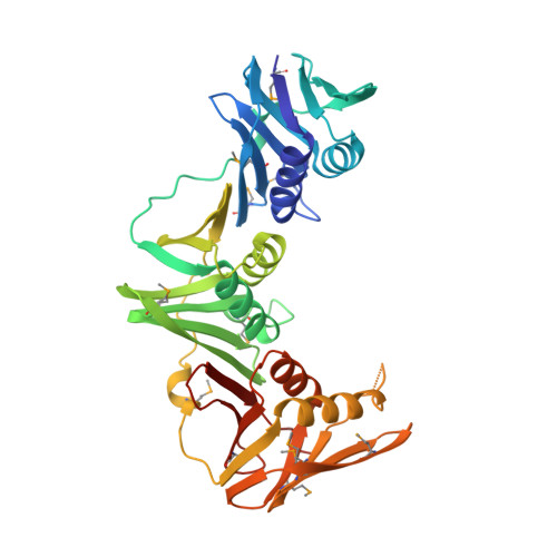 | |
UniProt | |||||
Entity Groups | |||||
| Sequence Clusters | 30% Identity50% Identity70% Identity90% Identity95% Identity100% Identity | ||||
| UniProt Group | C4Z938 | ||||
Sequence AnnotationsExpand | |||||
Reference Sequence | |||||
| Ligands 4 Unique | |||||
|---|---|---|---|---|---|
| ID | Chains | Name / Formula / InChI Key | 2D Diagram | 3D Interactions | |
| PGR Download:Ideal Coordinates CCD File | C [auth A], R [auth B] | R-1,2-PROPANEDIOL C3 H8 O2 DNIAPMSPPWPWGF-GSVOUGTGSA-N |  | ||
| EDO Download:Ideal Coordinates CCD File | AA [auth B] BA [auth B] F [auth A] G [auth A] H [auth A] | 1,2-ETHANEDIOL C2 H6 O2 LYCAIKOWRPUZTN-UHFFFAOYSA-N |  | ||
| ACT Download:Ideal Coordinates CCD File | D [auth A], E [auth A], S [auth B], T [auth B], U [auth B] | ACETATE ION C2 H3 O2 QTBSBXVTEAMEQO-UHFFFAOYSA-M |  | ||
| CL Download:Ideal Coordinates CCD File | CA [auth B], Q [auth A] | CHLORIDE ION Cl VEXZGXHMUGYJMC-UHFFFAOYSA-M |  | ||
| Modified Residues 1 Unique | |||||
|---|---|---|---|---|---|
| ID | Chains | Type | Formula | 2D Diagram | Parent |
| MSE Query on MSE | A, B | L-PEPTIDE LINKING | C5 H11 N O2 Se |  | MET |
| Length ( Å ) | Angle ( ˚ ) |
|---|---|
| a = 127.76 | α = 90 |
| b = 95.56 | β = 115.94 |
| c = 84.01 | γ = 90 |
| Software Name | Purpose |
|---|---|
| MolProbity | model building |
| PDB_EXTRACT | data extraction |
| SHELX | phasing |
| SHARP | phasing |
| XSCALE | data scaling |
| BUSTER-TNT | refinement |
| XDS | data reduction |
| SHELXD | phasing |
| BUSTER | refinement |


