3SZK

Crystal Structure of Human metHaemoglobin Complexed with the First NEAT Domain of IsdH from Staphylococcus aureus


Experimental Data Snapshot

  • Method: X-RAY DIFFRACTION
  • Resolution: 3.01 Å
  • R-Value Free: 0.275 
  • R-Value Work: 0.245 
  • R-Value Observed: 0.247 

Starting Models: experimental
View more details

wwPDB Validation   3D Report Full Report


Ligand Structure Quality Assessment 


This is version 1.2 of the entry. See complete history


Literature

Structural basis for hemoglobin capture by Staphylococcus aureus cell-surface protein, IsdH

Krishna Kumar, K.Jacques, D.A.Pishchany, G.Caradoc-Davies, T.Spirig, T.Malmirchegini, G.R.Langley, D.B.Dickson, C.F.Mackay, J.P.Clubb, R.T.Skaar, E.P.Guss, J.M.Gell, D.A.

(2011) J Biol Chem 286: 38439-38447

  • DOI: https://doi.org/10.1074/jbc.M111.287300
  • Primary Citation of Related Structures:  
    3SZK

  • PubMed Abstract: 

    Pathogens must steal iron from their hosts to establish infection. In mammals, hemoglobin (Hb) represents the largest reservoir of iron, and pathogens express Hb-binding proteins to access this source. Here, we show how one of the commonest and most significant human pathogens, Staphylococcus aureus, captures Hb as the first step of an iron-scavenging pathway. The x-ray crystal structure of Hb bound to a domain from the Isd (iron-regulated surface determinant) protein, IsdH, is the first structure of a Hb capture complex to be determined. Surface mutations in Hb that reduce binding to the Hb-receptor limit the capacity of S. aureus to utilize Hb as an iron source, suggesting that Hb sequence is a factor in host susceptibility to infection. The demonstration that pathogens make highly specific recognition complexes with Hb raises the possibility of developing inhibitors of Hb binding as antibacterial agents.


  • Organizational Affiliation

    School of Molecular Bioscience, University of Sydney, New South Wales 2006, Australia.


Macromolecules
Find similar proteins by:  (by identity cutoff)  |  3D Structure
Entity ID: 1
MoleculeChains Sequence LengthOrganismDetailsImage
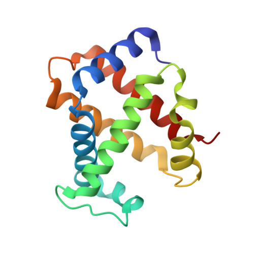
Hemoglobin subunit alphaA [auth D],
E [auth A]
141Homo sapiensMutation(s): 0 
UniProt & NIH Common Fund Data Resources
Find proteins for P69905 (Homo sapiens)
Explore P69905 
Go to UniProtKB:  P69905
PHAROS:  P69905
Entity Groups  
Sequence Clusters30% Identity50% Identity70% Identity90% Identity95% Identity100% Identity
UniProt GroupP69905
Sequence Annotations
Expand
  • Reference Sequence
Find similar proteins by:  (by identity cutoff)  |  3D Structure
Entity ID: 2
MoleculeChains Sequence LengthOrganismDetailsImage
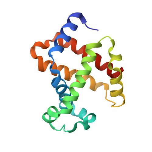
Hemoglobin subunit betaB,
C [auth E]
146Homo sapiensMutation(s): 0 
UniProt & NIH Common Fund Data Resources
Find proteins for P68871 (Homo sapiens)
Explore P68871 
Go to UniProtKB:  P68871
PHAROS:  P68871
GTEx:  ENSG00000244734 
Entity Groups  
Sequence Clusters30% Identity50% Identity70% Identity90% Identity95% Identity100% Identity
UniProt GroupP68871
Sequence Annotations
Expand
  • Reference Sequence
Find similar proteins by:  (by identity cutoff)  |  3D Structure
Entity ID: 3
MoleculeChains Sequence LengthOrganismDetailsImage
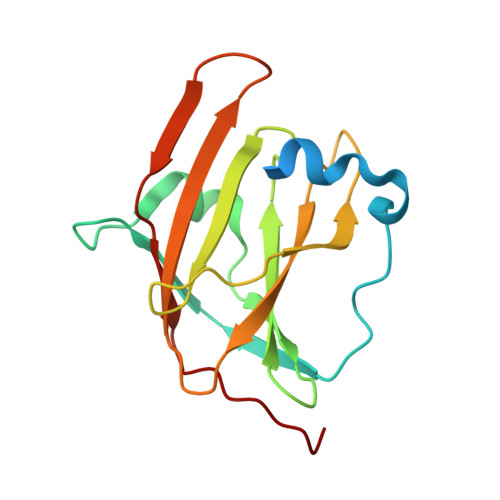
Iron-regulated surface determinant protein HD [auth F],
F [auth C]
164Staphylococcus aureus subsp. aureus MW2Mutation(s): 0 
UniProt
Find proteins for Q8NW39 (Staphylococcus aureus (strain MW2))
Explore Q8NW39 
Go to UniProtKB:  Q8NW39
Entity Groups  
Sequence Clusters30% Identity50% Identity70% Identity90% Identity95% Identity100% Identity
UniProt GroupQ8NW39
Sequence Annotations
Expand
  • Reference Sequence
Experimental Data & Validation

Experimental Data

  • Method: X-RAY DIFFRACTION
  • Resolution: 3.01 Å
  • R-Value Free: 0.275 
  • R-Value Work: 0.245 
  • R-Value Observed: 0.247 
  • Space Group: P 21 21 21
Unit Cell:
Length ( Å )Angle ( ˚ )
a = 65.88α = 90
b = 123.206β = 90
c = 143.933γ = 90
Software Package:
Software NamePurpose
HKL-2000data collection
PHASERphasing
REFMACrefinement
MOSFLMdata reduction
SCALEPACKdata scaling

Structure Validation

View Full Validation Report


Ligand Structure Quality Assessment 


Entry History 

Deposition Data

Revision History  (Full details and data files)

  • Version 1.0: 2011-09-14
    Type: Initial release
  • Version 1.1: 2012-05-09
    Changes: Database references
  • Version 1.2: 2023-11-01
    Changes: Data collection, Database references, Derived calculations, Refinement description