3SY2 | pdb_00003sy2

Crystal structure of the Salmonella E3 ubiquitin ligase SopA in complex with the human E2 UbcH7


Experimental Data Snapshot

  • Method: X-RAY DIFFRACTION
  • Resolution: 3.27 Å
  • R-Value Free: 
    0.275 (Depositor), 0.267 (DCC) 
  • R-Value Work: 
    0.211 (Depositor), 0.205 (DCC) 
  • R-Value Observed: 
    0.214 (Depositor) 

Starting Models: experimental
View more details

wwPDB Validation 3D Report Full Report

Validation slider image for 3SY2

This is version 1.2 of the entry. See complete history

Literature

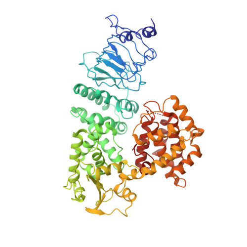
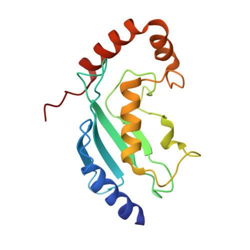
Crystal structures of two bacterial HECT-like E3 ligases in complex with a human E2 reveal atomic details of pathogen-host interactions.

Lin, D.Y.Diao, J.Chen, J.

(2012) Proc Natl Acad Sci U S A 109: 1925-1930

  • DOI: https://doi.org/10.1073/pnas.1115025109
  • Primary Citation Related Structures: 
    3SQV, 3SY2

  • PubMed Abstract: 

    In eukaryotes, ubiquitination is an important posttranslational process achieved through a cascade of ubiquitin-activating (E1), conjugating (E2), and ligase (E3) enzymes. Many pathogenic bacteria deliver virulence factors into the host cell that function as E3 ligases. How these bacterial "Trojan horses" integrate into the eukaryotic ubiquitin system has remained a mystery. Here we report crystal structures of two bacterial E3s, Salmonella SopA and Escherichia coli NleL, both in complex with human E2 UbcH7. These structures represent two distinct conformational states of the bacterial E3s, supporting the necessary structural rearrangements associated with ubiquitin transfer. The E2-interacting surface of SopA and NleL has little similarity to those of eukaryotic E3s. However, both bacterial E3s bind to the canonical surface of E2 that normally interacts with eukaryotic E3s. Furthermore, we show that a glutamate residue on E3 is involved in catalyzing ubiquitin transfer from E3 to the substrate, but not from E2 to E3. Together, these results provide mechanistic insights into the ubiquitin pathway and a framework for understanding molecular mimicry in bacterial pathogenesis.


  • Organizational Affiliation
    • Department of Biological Sciences, Purdue University, West Lafayette, IN 47907, USA.

Macromolecule Content 

  • Total Structure Weight: 174.63 kDa 
  • Atom Count: 11,660 
  • Modeled Residue Count: 1,533 
  • Deposited Residue Count: 1,554 
  • Unique protein chains: 2

Macromolecules

Find similar proteins by:|  3D Structure
Entity ID: 1
MoleculeChains  Sequence LengthOrganismDetailsImage
E3 ubiquitin-protein ligase SopA
A, B
621Salmonella enterica subsp. enterica serovar TyphimuriumMutation(s): 0 
Gene Names: sopASTM2066
EC: 6.3.2 (PDB Primary Data), 2.3.2.26 (UniProt)
UniProt
Find proteins for Q8ZNR3 (Salmonella typhimurium (strain LT2 / SGSC1412 / ATCC 700720))
Explore Q8ZNR3 
Go to UniProtKB:  Q8ZNR3
Entity Groups
Sequence Clusters30% Identity50% Identity70% Identity90% Identity95% Identity100% Identity
UniProt GroupQ8ZNR3
Sequence Annotations
Expand
Reference Sequence
Find similar proteins by:|  3D Structure
Entity ID: 2
MoleculeChains  Sequence LengthOrganismDetailsImage
Ubiquitin-conjugating enzyme E2 L3
C, D
156Homo sapiensMutation(s): 0 
Gene Names: UBE2L3UBCE7UBCH7
EC: 6.3.2.19 (PDB Primary Data), 2.3.2.23 (UniProt)
UniProt & NIH Common Fund Data Resources
Find proteins for P68036 (Homo sapiens)
Explore P68036 
Go to UniProtKB:  P68036
PHAROS:  P68036
GTEx:  ENSG00000185651 
Entity Groups
Sequence Clusters30% Identity50% Identity70% Identity90% Identity95% Identity100% Identity
UniProt GroupP68036
Sequence Annotations
Expand
Reference Sequence

Small Molecules

Ligands 1 Unique
IDChains Name / Formula / InChI Key2D Diagram3D Interactions
SO4

Query on SO4


Download:Ideal Coordinates CCD File
E [auth A]SULFATE ION
O4 S
QAOWNCQODCNURD-UHFFFAOYSA-L

Experimental Data & Validation

Experimental Data

  • Method: X-RAY DIFFRACTION
  • Resolution: 3.27 Å
  • R-Value Free:  0.275 (Depositor), 0.267 (DCC) 
  • R-Value Work:  0.211 (Depositor), 0.205 (DCC) 
  • R-Value Observed: 0.214 (Depositor) 
Space Group: P 21 21 21
Unit Cell:
Length ( Å )Angle ( ˚ )
a = 74.12α = 90
b = 118.363β = 90
c = 241.671γ = 90
Software Package:
Software NamePurpose
Blu-Icedata collection
PHASERphasing
PHENIXrefinement
HKL-2000data reduction
HKL-2000data scaling

Structure Validation

View Full Validation Report


Entry History 

Deposition Data

Revision History  (Full details and data files)

  • Version 1.0: 2012-01-25
    Type: Initial release
  • Version 1.1: 2012-02-22
    Changes: Database references
  • Version 1.2: 2023-09-13
    Changes: Data collection, Database references, Derived calculations, Refinement description