3SU8 | pdb_00003su8

Crystal structure of a truncated intracellular domain of Plexin-B1 in complex with Rac1


Experimental Data Snapshot

  • Method: X-RAY DIFFRACTION
  • Resolution: 3.20 Å
  • R-Value Free: 
    0.238 (Depositor), 0.227 (DCC) 
  • R-Value Work: 
    0.207 (Depositor) 
  • R-Value Observed: 
    0.208 (Depositor) 

wwPDB Validation 3D Report Full Report

Validation slider image for 3SU8

Ligand Structure Quality Assessment 


This is version 1.2 of the entry. See complete history

Literature

A Dual Binding Mode for RhoGTPases in Plexin Signalling.

Bell, C.H.Aricescu, A.R.Jones, E.Y.Siebold, C.

(2011) PLoS Biol 9: e1001134-e1001134

  • DOI: https://doi.org/10.1371/journal.pbio.1001134
  • Primary Citation Related Structures: 
    3SU8, 3SUA

  • PubMed Abstract: 

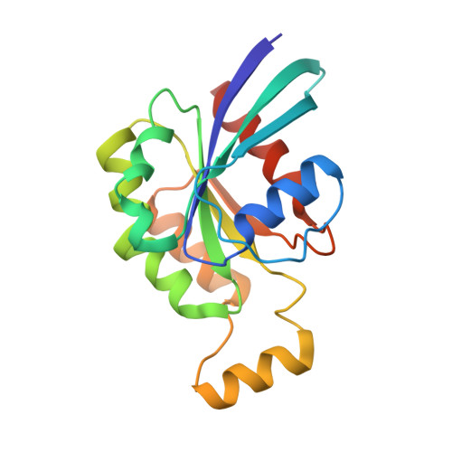
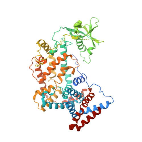
    Plexins are cell surface receptors for the semaphorin family of cell guidance cues. The cytoplasmic region comprises a Ras GTPase-activating protein (GAP) domain and a RhoGTPase binding domain. Concomitant binding of extracellular semaphorin and intracellular RhoGTPase triggers GAP activity and signal transduction. The mechanism of this intricate regulation remains elusive. We present two crystal structures of the human Plexin-B1 cytoplasmic region in complex with a constitutively active RhoGTPase, Rac1. The structure of truncated Plexin-B1-Rac1 complex provides no mechanism for coupling RhoGTPase and Ras binding sites. On inclusion of the juxtamembrane helix, a trimeric structure of Plexin-B1-Rac1 complexes is stabilised by a second, novel, RhoGTPase binding site adjacent to the Ras site. Site-directed mutagenesis combined with cellular and biophysical assays demonstrate that this new binding site is essential for signalling. Our findings are consistent with a model in which extracellular and intracellular plexin clustering events combine into a single signalling output.


  • Organizational Affiliation
    • Division of Structural Biology, Wellcome Trust Centre for Human Genetics, University of Oxford, Oxford, United Kingdom.

Macromolecule Content 

  • Total Structure Weight: 91.08 kDa 
  • Atom Count: 5,762 
  • Modeled Residue Count: 717 
  • Deposited Residue Count: 795 
  • Unique protein chains: 2

Macromolecules

Find similar proteins by:|  3D Structure
Entity ID: 1
MoleculeChains  Sequence LengthOrganismDetailsImage
Ras-related C3 botulinum toxin substrate 1184Homo sapiensMutation(s): 0 
Gene Names: RAC1TC25MIG5
EC: 3.6.5.2
UniProt & NIH Common Fund Data Resources
Find proteins for P63000 (Homo sapiens)
Explore P63000 
Go to UniProtKB:  P63000
PHAROS:  P63000
GTEx:  ENSG00000136238 
Entity Groups
Sequence Clusters30% Identity50% Identity70% Identity90% Identity95% Identity100% Identity
UniProt GroupP63000
Sequence Annotations
Expand
Reference Sequence
Find similar proteins by:|  3D Structure
Entity ID: 2
MoleculeChains  Sequence LengthOrganismDetailsImage
Plexin-B1B [auth X]611Homo sapiensMutation(s): 0 
Gene Names: PLXNB1KIAA0407PLXN5SEP
UniProt & NIH Common Fund Data Resources
Find proteins for O43157 (Homo sapiens)
Explore O43157 
Go to UniProtKB:  O43157
PHAROS:  O43157
GTEx:  ENSG00000164050 
Entity Groups
Sequence Clusters30% Identity50% Identity70% Identity90% Identity95% Identity100% Identity
UniProt GroupO43157
Sequence Annotations
Expand
Reference Sequence

Small Molecules

Ligands 2 Unique
IDChains Name / Formula / InChI Key2D Diagram3D Interactions
GNP

Query on GNP


Download:Ideal Coordinates CCD File
D [auth A]PHOSPHOAMINOPHOSPHONIC ACID-GUANYLATE ESTER
C10 H17 N6 O13 P3
UQABYHGXWYXDTK-UUOKFMHZSA-N
MG

Query on MG


Download:Ideal Coordinates CCD File
C [auth A]MAGNESIUM ION
Mg
JLVVSXFLKOJNIY-UHFFFAOYSA-N

Experimental Data & Validation

Experimental Data

  • Method: X-RAY DIFFRACTION
  • Resolution: 3.20 Å
  • R-Value Free:  0.238 (Depositor), 0.227 (DCC) 
  • R-Value Work:  0.207 (Depositor) 
  • R-Value Observed: 0.208 (Depositor) 
Space Group: C 1 2 1
Unit Cell:
Length ( Å )Angle ( ˚ )
a = 183.45α = 90
b = 63.81β = 107.53
c = 84.55γ = 90
Software Package:
Software NamePurpose
BUSTERrefinement

Structure Validation

View Full Validation Report


Ligand Structure Quality Assessment 


Entry History 

Deposition Data

Revision History  (Full details and data files)

  • Version 1.0: 2011-09-28
    Type: Initial release
  • Version 1.1: 2011-10-05
    Changes: Database references
  • Version 1.2: 2024-02-28
    Changes: Data collection, Database references, Derived calculations