3STM

Structure of human LFABP in complex with one molecule of palmitic acid


Experimental Data Snapshot

  • Method: X-RAY DIFFRACTION
  • Resolution: 2.22 Å
  • R-Value Free: 0.281 
  • R-Value Work: 0.242 
  • R-Value Observed: 0.243 

wwPDB Validation   3D Report Full Report


Ligand Structure Quality Assessment 


This is version 1.2 of the entry. See complete history


Literature

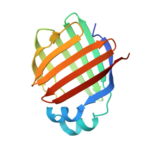
Fatty acid induced remodeling within the Human liver fatty acid binding protein

Sharma, A.Sharma, A.

(2011) J Biol Chem 

  • DOI: https://doi.org/10.1074/jbc.M111.270165
  • Primary Citation of Related Structures:  
    3STK, 3STM, 3STN

  • PubMed Abstract: 

    We crystallized human liver fatty acid-binding protein (LFABP) in apo, holo, and intermediate states of palmitic acid engagement. Structural snapshots of fatty acid recognition, entry, and docking within LFABP support a heads-in mechanism for ligand entry. Apo-LFABP undergoes structural remodeling, where the first palmitate ingress creates the atomic environment for placement of the second palmitate. These new mechanistic insights will facilitate development of pharmacological agents against LFABP.


  • Organizational Affiliation

    Structural and Computational Biology Group, International Centre for Genetic Engineering and Biotechnology, Aruna Asaf Ali Road, 110067 New Delhi, India.


Macromolecules
Find similar proteins by:  (by identity cutoff)  |  3D Structure
Entity ID: 1
MoleculeChains Sequence LengthOrganismDetailsImage
Fatty acid-binding protein, liverA [auth X]132Homo sapiensMutation(s): 0 
Gene Names: FABP1FABPL
UniProt & NIH Common Fund Data Resources
Find proteins for P07148 (Homo sapiens)
Explore P07148 
Go to UniProtKB:  P07148
PHAROS:  P07148
GTEx:  ENSG00000163586 
Entity Groups  
Sequence Clusters30% Identity50% Identity70% Identity90% Identity95% Identity100% Identity
UniProt GroupP07148
Sequence Annotations
Expand
  • Reference Sequence
Small Molecules
Ligands 1 Unique
IDChains Name / Formula / InChI Key2D Diagram3D Interactions
PLM
Query on PLM

Download Ideal Coordinates CCD File 
B [auth X]PALMITIC ACID
C16 H32 O2
IPCSVZSSVZVIGE-UHFFFAOYSA-N
Experimental Data & Validation

Experimental Data

  • Method: X-RAY DIFFRACTION
  • Resolution: 2.22 Å
  • R-Value Free: 0.281 
  • R-Value Work: 0.242 
  • R-Value Observed: 0.243 
  • Space Group: P 21 21 21
Unit Cell:
Length ( Å )Angle ( ˚ )
a = 30.236α = 90
b = 57.392β = 90
c = 74.404γ = 90
Software Package:
Software NamePurpose
SCALEPACKdata scaling
MOLREPphasing
PHENIXrefinement
PDB_EXTRACTdata extraction
MAR345dtbdata collection
DENZOdata reduction
HKL-2000data scaling

Structure Validation

View Full Validation Report


Ligand Structure Quality Assessment 


Entry History 

Deposition Data

Revision History  (Full details and data files)

  • Version 1.0: 2011-08-10
    Type: Initial release
  • Version 1.1: 2017-11-08
    Changes: Refinement description
  • Version 1.2: 2024-03-20
    Changes: Data collection, Database references, Derived calculations