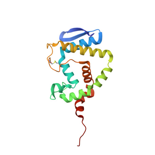
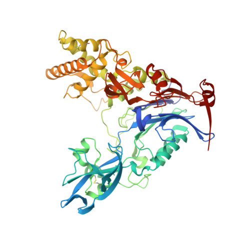
3SRA

Structure of Pseudomonas aerugionsa PvdQ covalently acylated with myristic acid from PVDIq


Experimental Data Snapshot

  • Method: X-RAY DIFFRACTION
  • Resolution: 2.30 Å
  • R-Value Free: 0.217 
  • R-Value Work: 0.182 
  • R-Value Observed: 0.184 

Starting Model: experimental
View more details

wwPDB Validation   3D Report Full Report


Ligand Structure Quality Assessment 


This is version 2.2 of the entry. See complete history


Literature

Structural Characterization and High-Throughput Screening of Inhibitors of PvdQ, an NTN Hydrolase Involved in Pyoverdine Synthesis.

Drake, E.J.Gulick, A.M.

(2011) ACS Chem Biol 6: 1277-1286

  • DOI: https://doi.org/10.1021/cb2002973
  • Primary Citation of Related Structures:  
    3L91, 3L94, 3SRA, 3SRB, 3SRC

  • PubMed Abstract: 

    The human pathogen Pseudomonas aeruginosa produces a variety of virulence factors including pyoverdine, a nonribosomally produced peptide siderophore. The maturation pathway of the pyoverdine peptide is complex and provides a unique target for inhibition. Within the pyoverdine biosynthetic cluster is a periplasmic hydrolase, PvdQ, that is required for pyoverdine production. However, the precise role of PvdQ in the maturation pathway has not been biochemically characterized. We demonstrate herein that the initial module of the nonribosomal peptide synthetase PvdL adds a myristate moiety to the pyoverdine precursor. We extracted this acylated precursor, called PVDIq, from a pvdQ mutant strain and show that the PvdQ enzyme removes the fatty acid catalyzing one of the final steps in pyoverdine maturation. Incubation of PVDIq with crystals of PvdQ allowed us to capture the acylated enzyme and confirm through structural studies the chemical composition of the incorporated acyl chain. Finally, because inhibition of siderophore synthesis has been identified as a potential antibiotic strategy, we developed a high-throughput screening assay and tested a small chemical library for compounds that inhibit PvdQ activity. Two compounds that block PvdQ have been identified, and their binding within the fatty acid binding pocket was structurally characterized.


  • Organizational Affiliation

    Hauptman-Woodward Medical Research Institute and Department of Structural Biology, State University of New York at Buffalo, 700 Ellicott Street, Buffalo, New York 14203-1102, United States.


Macromolecules
Find similar proteins by:  (by identity cutoff)  |  3D Structure
Entity ID: 1
MoleculeChains Sequence LengthOrganismDetailsImage
Acyl-homoserine lactone acylase PvdQ subunit alpha163Pseudomonas aeruginosa PAO1Mutation(s): 0 
Gene Names: pvdQqsc112PA2385
EC: 3.5.1.97
UniProt
Find proteins for Q9I194 (Pseudomonas aeruginosa (strain ATCC 15692 / DSM 22644 / CIP 104116 / JCM 14847 / LMG 12228 / 1C / PRS 101 / PAO1))
Explore Q9I194 
Go to UniProtKB:  Q9I194
Entity Groups  
Sequence Clusters30% Identity50% Identity70% Identity90% Identity95% Identity100% Identity
UniProt GroupQ9I194
Sequence Annotations
Expand
  • Reference Sequence
Find similar proteins by:  (by identity cutoff)  |  3D Structure
Entity ID: 2
MoleculeChains Sequence LengthOrganismDetailsImage
Acyl-homoserine lactone acylase PvdQ subunit beta546Pseudomonas aeruginosa PAO1Mutation(s): 0 
Gene Names: pvdQqsc112PA2385
EC: 3.5.1.97
UniProt
Find proteins for Q9I194 (Pseudomonas aeruginosa (strain ATCC 15692 / DSM 22644 / CIP 104116 / JCM 14847 / LMG 12228 / 1C / PRS 101 / PAO1))
Explore Q9I194 
Go to UniProtKB:  Q9I194
Entity Groups  
Sequence Clusters30% Identity50% Identity70% Identity90% Identity95% Identity100% Identity
UniProt GroupQ9I194
Sequence Annotations
Expand
  • Reference Sequence
Small Molecules
Ligands 2 Unique
IDChains Name / Formula / InChI Key2D Diagram3D Interactions
MYR
Query on MYR

Download Ideal Coordinates CCD File 
V [auth B]MYRISTIC ACID
C14 H28 O2
TUNFSRHWOTWDNC-UHFFFAOYSA-N
EDO
Query on EDO

Download Ideal Coordinates CCD File 
C [auth A]
D [auth A]
E [auth A]
F [auth A]
G [auth A]
C [auth A],
D [auth A],
E [auth A],
F [auth A],
G [auth A],
H [auth A],
I [auth A],
J [auth A],
K [auth A],
L [auth B],
M [auth B],
N [auth B],
O [auth B],
P [auth B],
Q [auth B],
R [auth B],
S [auth B],
T [auth B],
U [auth B]
1,2-ETHANEDIOL
C2 H6 O2
LYCAIKOWRPUZTN-UHFFFAOYSA-N
Experimental Data & Validation

Experimental Data

  • Method: X-RAY DIFFRACTION
  • Resolution: 2.30 Å
  • R-Value Free: 0.217 
  • R-Value Work: 0.182 
  • R-Value Observed: 0.184 
  • Space Group: C 2 2 21
Unit Cell:
Length ( Å )Angle ( ˚ )
a = 120.168α = 90
b = 166.622β = 90
c = 93.746γ = 90
Software Package:
Software NamePurpose
Blu-Icedata collection
MOLREPphasing
REFMACrefinement
HKL-2000data reduction
HKL-2000data scaling

Structure Validation

View Full Validation Report


Ligand Structure Quality Assessment 


Entry History 

Deposition Data

Revision History  (Full details and data files)

  • Version 1.0: 2011-09-21
    Type: Initial release
  • Version 1.1: 2011-11-30
    Changes: Database references
  • Version 2.0: 2023-05-31
    Changes: Atomic model, Database references, Derived calculations, Source and taxonomy, Structure summary
  • Version 2.1: 2023-09-20
    Changes: Data collection, Refinement description
  • Version 2.2: 2024-10-30
    Changes: Structure summary