3SOV | pdb_00003sov

The structure of a beta propeller domain in complex with peptide S


Experimental Data Snapshot

  • Method: X-RAY DIFFRACTION
  • Resolution: 1.27 Å
  • R-Value Free: 
    0.180 (Depositor), 0.159 (DCC) 
  • R-Value Work: 
    0.153 (Depositor), 0.157 (DCC) 
  • R-Value Observed: 
    0.154 (Depositor) 

wwPDB Validation 3D Report Full Report

Validation slider image for 3SOV

Ligand Structure Quality Assessment 


This is version 1.3 of the entry. See complete history

Literature

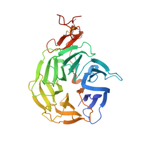
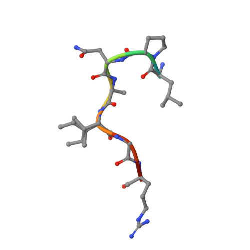
Wnt antagonists bind through a short peptide to the first beta-propeller domain of LRP5/6.

Bourhis, E.Wang, W.Tam, C.Hwang, J.Zhang, Y.Spittler, D.Huang, O.W.Gong, Y.Estevez, A.Zilberleyb, I.Rouge, L.Chiu, C.Wu, Y.Costa, M.Hannoush, R.N.Franke, Y.Cochran, A.G.

(2011) Structure 19: 1433-1442

  • DOI: https://doi.org/10.1016/j.str.2011.07.005
  • Primary Citation Related Structures: 
    3SOB, 3SOQ, 3SOV

  • PubMed Abstract: 

    The Wnt pathway inhibitors DKK1 and sclerostin (SOST) are important therapeutic targets in diseases involving bone loss or damage. It has been appreciated that Wnt coreceptors LRP5/6 are also important, as human missense mutations that result in bone overgrowth (bone mineral density, or BMD, mutations) cluster to the E1 propeller domain of LRP5. Here, we report a crystal structure of LRP6 E1 bound to an antibody, revealing that the E1 domain is a peptide recognition module. Remarkably, the consensus E1 binding sequence is a close match to a conserved tripeptide motif present in all Wnt inhibitors that bind LRP5/6. We show that this motif is important for DKK1 and SOST binding to LRP6 and for inhibitory function, providing a detailed structural explanation for the effect of the BMD mutations.


  • Organizational Affiliation
    • Department of Early Discovery Biochemistry, Genentech Research and Early Development, 1 DNA Way, South San Francisco, CA 94080, USA.

Macromolecule Content 

  • Total Structure Weight: 37.83 kDa 
  • Atom Count: 3,050 
  • Modeled Residue Count: 313 
  • Deposited Residue Count: 327 
  • Unique protein chains: 2

Macromolecules

Find similar proteins by:|  3D Structure
Entity ID: 1
MoleculeChains  Sequence LengthOrganismDetailsImage
Low-density lipoprotein receptor-related protein 6318Homo sapiensMutation(s): 0 
Gene Names: LRP6
UniProt & NIH Common Fund Data Resources
Find proteins for O75581 (Homo sapiens)
Explore O75581 
Go to UniProtKB:  O75581
PHAROS:  O75581
GTEx:  ENSG00000070018 
Entity Groups
Sequence Clusters30% Identity50% Identity70% Identity90% Identity95% Identity100% Identity
UniProt GroupO75581
Sequence Annotations
Expand
Reference Sequence
Find similar proteins by:  Sequence   |   3D Structure  
Entity ID: 2
MoleculeChains  Sequence LengthOrganismDetailsImage
SclerostinB [auth Z]9Homo sapiensMutation(s): 2 
UniProt & NIH Common Fund Data Resources
Find proteins for Q9BQB4 (Homo sapiens)
Explore Q9BQB4 
Go to UniProtKB:  Q9BQB4
PHAROS:  Q9BQB4
GTEx:  ENSG00000167941 
Entity Groups
UniProt GroupQ9BQB4
Sequence Annotations
Expand
Reference Sequence

Small Molecules

Ligands 3 Unique
IDChains Name / Formula / InChI Key2D Diagram3D Interactions
NAG

Query on NAG


Download:Ideal Coordinates CCD File
C [auth A],
G [auth A]
2-acetamido-2-deoxy-beta-D-glucopyranose
C8 H15 N O6
OVRNDRQMDRJTHS-FMDGEEDCSA-N
FUC

Query on FUC


Download:Ideal Coordinates CCD File
D [auth A],
E [auth A],
F [auth A],
H [auth A]
alpha-L-fucopyranose
C6 H12 O5
SHZGCJCMOBCMKK-SXUWKVJYSA-N
GOL

Query on GOL


Download:Ideal Coordinates CCD File
I [auth A],
J [auth A],
K [auth A]
GLYCEROL
C3 H8 O3
PEDCQBHIVMGVHV-UHFFFAOYSA-N

Experimental Data & Validation

Experimental Data

  • Method: X-RAY DIFFRACTION
  • Resolution: 1.27 Å
  • R-Value Free:  0.180 (Depositor), 0.159 (DCC) 
  • R-Value Work:  0.153 (Depositor), 0.157 (DCC) 
  • R-Value Observed: 0.154 (Depositor) 
Space Group: C 1 2 1
Unit Cell:
Length ( Å )Angle ( ˚ )
a = 103.567α = 90
b = 47.09β = 97.68
c = 68.72γ = 90
Software Package:
Software NamePurpose
HKL-2000data collection
PHASERphasing
PHENIXrefinement
HKL-2000data reduction
HKL-2000data scaling

Structure Validation

View Full Validation Report


Ligand Structure Quality Assessment 


Entry History 

Deposition Data

Revision History  (Full details and data files)

  • Version 1.0: 2011-09-21
    Type: Initial release
  • Version 1.1: 2013-08-28
    Changes: Database references
  • Version 1.2: 2020-07-29
    Type: Remediation
    Reason: Carbohydrate remediation
    Changes: Data collection, Database references, Derived calculations, Structure summary
  • Version 1.3: 2024-10-16
    Changes: Data collection, Database references, Structure summary