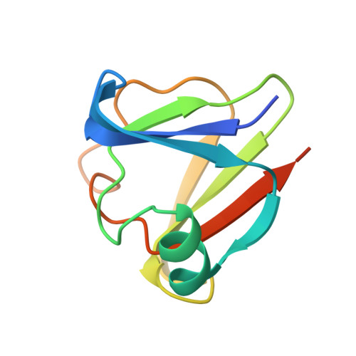
Decoding the molecular design principles underlying Ca(2+) binding to beta gamma-crystallin motifs
Mishra, A., Suman, S.K., Srivastava, S.S., Sankaranarayanan, R., Sharma, Y.(2012) J Mol Biol 415: 75-91
- PubMed: 22099475 Search on PubMed
- DOI: https://doi.org/10.1016/j.jmb.2011.10.037
- Primary Citation Related Structures:
3SNY, 3SNZ, 3SO0, 3SO1 - PubMed Abstract:
Numerous proteins belonging to the recently expanded βγ-crystallin superfamily bind Ca(2+) at the double-clamp N/D-N/D-X(1)-X(2)-S/T-S motif. However, there have been no attempts to understand the intricacies involving Ca(2+) binding, such as the determinants of Ca(2+)-binding affinity and their contributions to gain in stability. This work is an in-depth analysis of understanding the modes and determinants of Ca(2+) binding to βγ-crystallin motifs. We have performed extensive naturally occurring substitutions from related proteins on the βγ-crystallin domains of flavollin, a low-affinity Ca(2+)-binding protein, and clostrillin, a moderate-affinity protein. We monitored the consequences of these modifications on Ca(2)(+) binding by isothermal titration calorimetry, thermal stability and conformational and crystal structure analyses. We demonstrate that Ca(2)(+) binding to the two sites of a βγ-domain is interdependent and that the presence of Arg at the fifth position disables a site. A change from Thr to Ser, or vice versa, influences Ca(2+)-binding affinity, highlighting the basis of diversity found in these domains. A subtle change in the first site has a greater influence on Ca(2)(+) binding than a similar alteration in the second site. Thus, the second site is more variable in nature. Replacing an acidic or hydrophobic residue in a binding site alters the Ca(2+)-binding properties drastically. While it appears from their binding site sequence that these domains have evolved randomly, our examination illustrates the subtlety in the design of these modules. Decoding such design schemes would aid in our understanding of the functional themes underlying differential Ca(2)(+) binding and in predicting these in emerging sequence information.
- Centre for Cellular and Molecular Biology, Council of Scientific and Industrial Research, Hyderabad 500007, India.
Organizational Affiliation: