


Crystal structure of BA2930 in complex with AcCoA and cytosine
Klimecka, M.M., Chruszcz, M., Porebski, P.J., Cymborowski, M., Anderson, W., Minor, W.To be published.
Experimental Data Snapshot
Starting Model: experimental
View more details
Entity ID: 1 | |||||
|---|---|---|---|---|---|
| Molecule | Chains | Sequence Length | Organism | Details | Image |
| Aminoglycoside N3-acetyltransferase | A, B [auth D], C, D [auth B] | 268 | Bacillus anthracis | Mutation(s): 1 Gene Names: aacC7, BA_2930, GBAA2930, GBAA_2930 EC: 2.3.1.81 (PDB Primary Data), 2.3.1 (UniProt) | 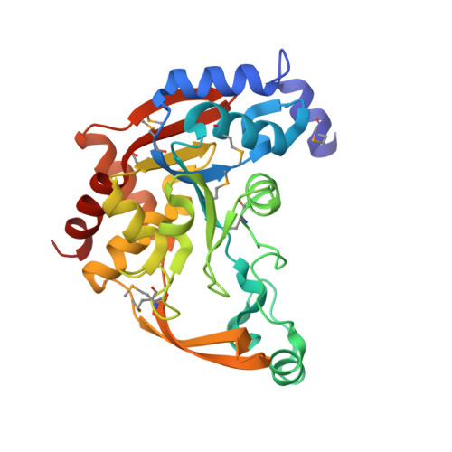 |
UniProt | |||||
Entity Groups | |||||
| Sequence Clusters | 30% Identity50% Identity70% Identity90% Identity95% Identity100% Identity | ||||
| UniProt Group | A0A3P1UCA6 | ||||
Sequence AnnotationsExpand | |||||
Reference Sequence | |||||
| Ligands 5 Unique | |||||
|---|---|---|---|---|---|
| ID | Chains | Name / Formula / InChI Key | 2D Diagram | 3D Interactions | |
| ACO Download:Ideal Coordinates CCD File | E [auth A], H [auth D], M [auth C], P [auth B] | ACETYL COENZYME *A C23 H38 N7 O17 P3 S ZSLZBFCDCINBPY-ZSJPKINUSA-N |  | ||
| CPS Download:Ideal Coordinates CCD File | O [auth C] | 3-[(3-CHOLAMIDOPROPYL)DIMETHYLAMMONIO]-1-PROPANESULFONATE C32 H58 N2 O7 S UMCMPZBLKLEWAF-BCTGSCMUSA-N |  | ||
| EPE Download:Ideal Coordinates CCD File | K [auth D] | 4-(2-HYDROXYETHYL)-1-PIPERAZINE ETHANESULFONIC ACID C8 H18 N2 O4 S JKMHFZQWWAIEOD-UHFFFAOYSA-N |  | ||
| CYT Download:Ideal Coordinates CCD File | F [auth A], I [auth D], N [auth C], Q [auth B] | 6-AMINOPYRIMIDIN-2(1H)-ONE C4 H5 N3 O OPTASPLRGRRNAP-UHFFFAOYSA-N |  | ||
| MG Download:Ideal Coordinates CCD File | G [auth A], J [auth D], L [auth D], R [auth B] | MAGNESIUM ION Mg JLVVSXFLKOJNIY-UHFFFAOYSA-N |  | ||
| Modified Residues 1 Unique | |||||
|---|---|---|---|---|---|
| ID | Chains | Type | Formula | 2D Diagram | Parent |
| MSE Query on MSE | A, B [auth D], C, D [auth B] | L-PEPTIDE LINKING | C5 H11 N O2 Se |  | MET |
| Length ( Å ) | Angle ( ˚ ) |
|---|---|
| a = 72.473 | α = 90 |
| b = 110.63 | β = 112.24 |
| c = 73.78 | γ = 90 |
| Software Name | Purpose |
|---|---|
| MD2 | data collection |
| HKL-3000 | phasing |
| MOLREP | phasing |
| REFMAC | refinement |
| Coot | model building |
| HKL-3000 | data reduction |
| HKL-3000 | data scaling |


