3SHS

Three N-terminal domains of the bacteriophage RB49 Highly Immunogenic Outer Capsid protein (Hoc)


Experimental Data Snapshot

  • Method: X-RAY DIFFRACTION
  • Resolution: 1.95 Å
  • R-Value Free: 0.267 
  • R-Value Work: 0.218 
  • R-Value Observed: 0.223 

wwPDB Validation   3D Report Full Report


This is version 1.4 of the entry. See complete history


Literature

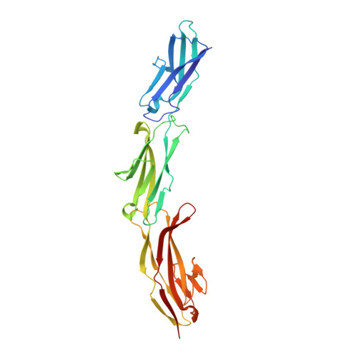
Structure of the three N-terminal immunoglobulin domains of the highly immunogenic outer capsid protein from a T4-like bacteriophage.

Fokine, A.Islam, M.Z.Zhang, Z.Bowman, V.D.Rao, V.B.Rossmann, M.G.

(2011) J Virol 85: 8141-8148

  • DOI: https://doi.org/10.1128/JVI.00847-11
  • Primary Citation of Related Structures:  
    3SHS

  • PubMed Abstract: 

    The head of bacteriophage T4 is decorated with 155 copies of the highly antigenic outer capsid protein (Hoc). One Hoc molecule binds near the center of each hexameric capsomer. Hoc is dispensable for capsid assembly and has been used to display pathogenic antigens on the surface of T4. Here we report the crystal structure of a protein containing the first three of four domains of Hoc from bacteriophage RB49, a close relative of T4. The structure shows an approximately linear arrangement of the protein domains. Each of these domains has an immunoglobulin-like fold, frequently found in cell attachment molecules. In addition, we report biochemical data suggesting that Hoc can bind to Escherichia coli, supporting the hypothesis that Hoc could attach the phage capsids to bacterial surfaces and perhaps also to other organisms. The capacity for such reversible adhesion probably provides survival advantages to the bacteriophage.


  • Organizational Affiliation

    Department of Biological Sciences, Purdue University, West Lafayette, Indiana 47907-2032, USA.


Macromolecules
Find similar proteins by:  (by identity cutoff)  |  3D Structure
Entity ID: 1
MoleculeChains Sequence LengthOrganismDetailsImage
Hoc head outer capsid protein304Escherichia phage RB49Mutation(s): 0 
Gene Names: hoc
UniProt
Find proteins for Q7Y442 (Escherichia phage RB49)
Explore Q7Y442 
Go to UniProtKB:  Q7Y442
Entity Groups  
Sequence Clusters30% Identity50% Identity70% Identity90% Identity95% Identity100% Identity
UniProt GroupQ7Y442
Sequence Annotations
Expand
  • Reference Sequence
Small Molecules
Ligands 1 Unique
IDChains Name / Formula / InChI Key2D Diagram3D Interactions
MG
Query on MG

Download Ideal Coordinates CCD File 
B [auth A]MAGNESIUM ION
Mg
JLVVSXFLKOJNIY-UHFFFAOYSA-N
Experimental Data & Validation

Experimental Data

  • Method: X-RAY DIFFRACTION
  • Resolution: 1.95 Å
  • R-Value Free: 0.267 
  • R-Value Work: 0.218 
  • R-Value Observed: 0.223 
  • Space Group: C 1 2 1
Unit Cell:
Length ( Å )Angle ( ˚ )
a = 213.54α = 90
b = 36.731β = 99.28
c = 62.693γ = 90
Software Package:
Software NamePurpose
Blu-Icedata collection
PHENIXmodel building
SOLVEphasing
PHENIXrefinement
HKL-2000data reduction
HKL-2000data scaling
PHENIXphasing

Structure Validation

View Full Validation Report


Entry History 

Deposition Data

Revision History  (Full details and data files)

  • Version 1.0: 2011-06-29
    Type: Initial release
  • Version 1.1: 2011-07-13
    Changes: Version format compliance
  • Version 1.2: 2011-11-30
    Changes: Database references
  • Version 1.3: 2017-11-08
    Changes: Refinement description
  • Version 1.4: 2024-02-28
    Changes: Data collection, Database references, Derived calculations