3SH0

Crystal Structure of E. coli undecaprenyl pyrophosphate synthase in complex with BPH-1065


Experimental Data Snapshot

  • Method: X-RAY DIFFRACTION
  • Resolution: 1.84 Å
  • R-Value Free: 0.211 
  • R-Value Work: 0.168 
  • R-Value Observed: 0.170 

wwPDB Validation   3D Report Full Report


Ligand Structure Quality Assessment 


This is version 1.2 of the entry. See complete history


Literature

Antibacterial drug leads targeting isoprenoid biosynthesis.

Zhu, W.Zhang, Y.Sinko, W.Hensler, M.E.Olson, J.Molohon, K.J.Lindert, S.Cao, R.Li, K.Wang, K.Wang, Y.Liu, Y.L.Sankovsky, A.de Oliveira, C.A.Mitchell, D.A.Nizet, V.McCammon, J.A.Oldfield, E.

(2013) Proc Natl Acad Sci U S A 110: 123-128

  • DOI: https://doi.org/10.1073/pnas.1219899110
  • Primary Citation of Related Structures:  
    3SGT, 3SGV, 3SGX, 3SH0, 4H2J, 4H2M, 4H2O, 4H38, 4H3A, 4H3C, 4H8E

  • PubMed Abstract: 

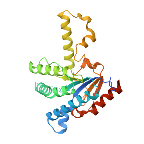
    With the rise in resistance to antibiotics such as methicillin, there is a need for new drugs. We report here the discovery and X-ray crystallographic structures of 10 chemically diverse compounds (benzoic, diketo, and phosphonic acids, as well as a bisamidine and a bisamine) that inhibit bacterial undecaprenyl diphosphate synthase, an essential enzyme involved in cell wall biosynthesis. The inhibitors bind to one or more of the four undecaprenyl diphosphate synthase inhibitor binding sites identified previously, with the most active leads binding to site 4, outside the catalytic center. The most potent leads are active against Staphylococcus aureus [minimal inhibitory concentration (MIC)(90) ∼0.25 µg/mL], and one potently synergizes with methicillin (fractional inhibitory concentration index = 0.25) and is protective in a mouse infection model. These results provide numerous leads for antibacterial development and open up the possibility of restoring sensitivity to drugs such as methicillin, using combination therapies.


  • Organizational Affiliation

    Center for Biophysics and Computational Biology, University of Illinois, Urbana, IL 61801, USA.


Macromolecules
Find similar proteins by:  (by identity cutoff)  |  3D Structure
Entity ID: 1
MoleculeChains Sequence LengthOrganismDetailsImage
Undecaprenyl pyrophosphate synthaseA [auth B],
B [auth A]
253Escherichia coli K-12Mutation(s): 0 
Gene Names: uppSispUrthyaeSb0174JW0169
EC: 2.5.1.31
UniProt
Find proteins for P60472 (Escherichia coli (strain K12))
Explore P60472 
Go to UniProtKB:  P60472
Entity Groups  
Sequence Clusters30% Identity50% Identity70% Identity90% Identity95% Identity100% Identity
UniProt GroupP60472
Sequence Annotations
Expand
  • Reference Sequence
Small Molecules
Binding Affinity Annotations 
IDSourceBinding Affinity
SAX PDBBind:  3SH0 IC50: 2200 (nM) from 1 assay(s)
Experimental Data & Validation

Experimental Data

  • Method: X-RAY DIFFRACTION
  • Resolution: 1.84 Å
  • R-Value Free: 0.211 
  • R-Value Work: 0.168 
  • R-Value Observed: 0.170 
  • Space Group: P 21 21 21
Unit Cell:
Length ( Å )Angle ( ˚ )
a = 62.765α = 90
b = 68.724β = 90
c = 112.056γ = 90
Software Package:
Software NamePurpose
SCALEPACKdata scaling
REFMACrefinement
PDB_EXTRACTdata extraction
HKL-2000data collection
HKL-2000data reduction
HKL-2000data scaling
PHASERphasing

Structure Validation

View Full Validation Report


Ligand Structure Quality Assessment 


Entry History 

Deposition Data

Revision History  (Full details and data files)

  • Version 1.0: 2012-12-19
    Type: Initial release
  • Version 1.1: 2013-01-16
    Changes: Database references
  • Version 1.2: 2023-09-13
    Changes: Data collection, Database references, Derived calculations, Refinement description