3SEL

PpcA M58N mutant


Experimental Data Snapshot

  • Method: X-RAY DIFFRACTION
  • Resolution: 2.10 Å
  • R-Value Free: 0.223 
  • R-Value Work: 0.176 
  • R-Value Observed: 0.180 

wwPDB Validation   3D Report Full Report


Ligand Structure Quality Assessment 


This is version 2.1 of the entry. See complete history


Literature

Pitfalls in the interpretation of structural changes in mutant proteins from crystal structures.

Pokkuluri, P.R.Yang, X.Londer, Y.Y.Schiffer, M.

(2012) J Struct Funct Genomics 13: 227-232

  • DOI: https://doi.org/10.1007/s10969-012-9147-1
  • Primary Citation of Related Structures:  
    3SEL, 3SJ0, 3SJ1, 3SJ4

  • PubMed Abstract: 

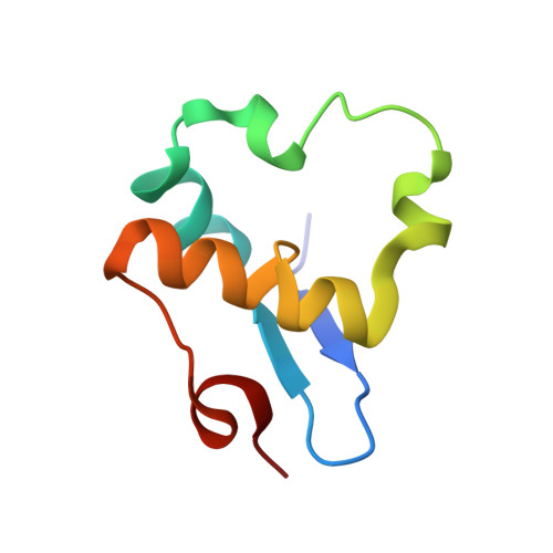
    PpcA is a small protein with 71 residues that contains three covalently bound hemes. The structures of single mutants at residue 58 have shown larger deviations in another part of the protein molecule than at the site of the mutation. Closer examination of the crystal packing has revealed the origin of this unexpected structural change. The site of mutation is within Van der Waals distance from another protein molecule related by a crystallographic twofold axis within the crystal. The structural changes occurred at or near the mutation site have led to a slight adjustment of the surface residues in contact. The observed deviations between the native and the mutant molecular structures are derived from the new crystal packing even though the two crystals are essentially isomorphous. Without careful consideration of the crystal lattice a non-expert looking at only the coordinates deposited in the Protein Data Bank could draw erroneous conclusion that mutation in one part of the molecule affected the structure of the protein in a distant part of the molecule.


  • Organizational Affiliation

    Biosciences Division, Argonne National Laboratory, Lemont, IL 60439, USA.


Macromolecules
Find similar proteins by:  (by identity cutoff)  |  3D Structure
Entity ID: 1
MoleculeChains Sequence LengthOrganismDetailsImage
CYTOCHROME C7A [auth X]71Geobacter sulfurreducensMutation(s): 1 
Gene Names: ppcAGSU0612
UniProt
Find proteins for Q8GGK7 (Geobacter sulfurreducens)
Explore Q8GGK7 
Go to UniProtKB:  Q8GGK7
Entity Groups  
Sequence Clusters30% Identity50% Identity70% Identity90% Identity95% Identity100% Identity
UniProt GroupQ8GGK7
Sequence Annotations
Expand
  • Reference Sequence
Experimental Data & Validation

Experimental Data

  • Method: X-RAY DIFFRACTION
  • Resolution: 2.10 Å
  • R-Value Free: 0.223 
  • R-Value Work: 0.176 
  • R-Value Observed: 0.180 
  • Space Group: P 43 2 2
Unit Cell:
Length ( Å )Angle ( ˚ )
a = 32.42α = 90
b = 32.42β = 90
c = 177.45γ = 90
Software Package:
Software NamePurpose
REFMACrefinement
CNSrefinement
HKL-2000data collection
HKL-2000data reduction
HKL-2000data scaling
CNSphasing

Structure Validation

View Full Validation Report


Ligand Structure Quality Assessment 


Entry History 

Deposition Data

Revision History  (Full details and data files)

  • Version 1.0: 2012-06-27
    Type: Initial release
  • Version 1.1: 2012-12-19
    Changes: Database references
  • Version 1.2: 2019-07-17
    Changes: Advisory, Data collection, Derived calculations, Refinement description
  • Version 2.0: 2021-03-03
    Changes: Atomic model, Data collection, Database references, Derived calculations, Non-polymer description, Structure summary
  • Version 2.1: 2023-09-13
    Changes: Advisory, Data collection, Database references, Refinement description