3S70 | pdb_00003s70

Crystal structure of active caspase-6 bound with Ac-VEID-CHO solved by As-SAD


Experimental Data Snapshot

  • Method: X-RAY DIFFRACTION
  • Resolution: 1.63 Å
  • R-Value Free: 
    0.194 (Depositor), 0.194 (DCC) 
  • R-Value Work: 
    0.164 (Depositor), 0.164 (DCC) 
  • R-Value Observed: 
    0.165 (Depositor) 

wwPDB Validation 3D Report Full Report

Validation slider image for 3S70

This is version 1.2 of the entry. See complete history

Literature

Get phases from arsenic anomalous scattering: de novo SAD phasing of two protein structures crystallized in cacodylate buffer

Liu, X.Zhang, H.Wang, X.-J.Li, L.-F.Su, X.-D.

(2011) PLoS One 6: e24227-e24227

  • DOI: https://doi.org/10.1371/journal.pone.0024227
  • Primary Citation Related Structures: 
    3S2S, 3S70

  • PubMed Abstract: 

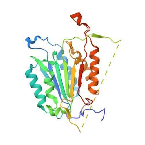
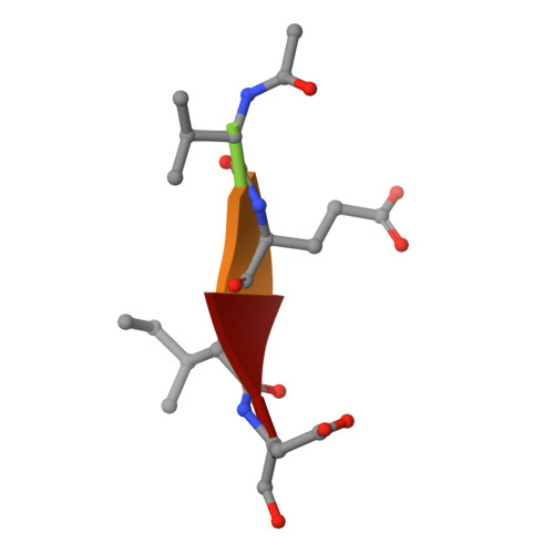
    The crystal structures of two proteins, a putative pyrazinamidase/nicotinamidase from the dental pathogen Streptococcus mutans (SmPncA) and the human caspase-6 (Casp6), were solved by de novo arsenic single-wavelength anomalous diffraction (As-SAD) phasing method. Arsenic (As), an uncommonly used element in SAD phasing, was covalently introduced into proteins by cacodylic acid, the buffering agent in the crystallization reservoirs. In SmPncA, the only cysteine was bound to dimethylarsinoyl, which is a pentavalent arsenic group (As (V)). This arsenic atom and a protein-bound zinc atom both generated anomalous signals. The predominant contribution, however, was from the As anomalous signals, which were sufficient to phase the SmPncA structure alone. In Casp6, four cysteines were found to bind cacodyl, a trivalent arsenic group (As (III)), in the presence of the reducing agent, dithiothreitol (DTT), and arsenic atoms were the only anomalous scatterers for SAD phasing. Analyses and discussion of these two As-SAD phasing examples and comparison of As with other traditional heavy atoms that generate anomalous signals, together with a few arsenic-based de novo phasing cases reported previously strongly suggest that As is an ideal anomalous scatterer for SAD phasing in protein crystallography.


  • Organizational Affiliation
    • State Key Laboratory of Protein and Plant Gene Research and Biodynamic Optical Imaging Center (BIOPIC), School of Life Sciences, Peking University, Beijing, China.

Macromolecule Content 

  • Total Structure Weight: 66.26 kDa 
  • Atom Count: 4,642 
  • Modeled Residue Count: 490 
  • Deposited Residue Count: 566 
  • Unique protein chains: 2

Macromolecules

Find similar proteins by:|  3D Structure
Entity ID: 1
MoleculeChains  Sequence LengthOrganismDetailsImage
Caspase-6
A, C
278Homo sapiensMutation(s): 0 
Gene Names: CASP6
EC: 3.4.22.59
UniProt & NIH Common Fund Data Resources
Find proteins for P55212 (Homo sapiens)
Explore P55212 
Go to UniProtKB:  P55212
PHAROS:  P55212
GTEx:  ENSG00000138794 
Entity Groups
Sequence Clusters30% Identity50% Identity70% Identity90% Identity95% Identity100% Identity
UniProt GroupP55212
Sequence Annotations
Expand
Reference Sequence
Find similar proteins by:  Sequence   |   3D Structure  
Entity ID: 2
MoleculeChains  Sequence LengthOrganismDetailsImage
aldehyde inhibitor Ac-VEID-CHO
B, D
5N/AMutation(s): 0 

Small Molecules

Ligands 3 Unique
IDChains Name / Formula / InChI Key2D Diagram3D Interactions
CAC

Query on CAC


Download:Ideal Coordinates CCD File
E [auth A]
F [auth A]
G [auth A]
H [auth A]
K [auth C]
E [auth A],
F [auth A],
G [auth A],
H [auth A],
K [auth C],
L [auth C],
M [auth C],
N [auth C]
CACODYLATE ION
C2 H6 As O2
OGGXGZAMXPVRFZ-UHFFFAOYSA-M
ACT

Query on ACT


Download:Ideal Coordinates CCD File
I [auth A]ACETATE ION
C2 H3 O2
QTBSBXVTEAMEQO-UHFFFAOYSA-M
MG

Query on MG


Download:Ideal Coordinates CCD File
J [auth A]MAGNESIUM ION
Mg
JLVVSXFLKOJNIY-UHFFFAOYSA-N
Modified Residues  1 Unique
IDChains TypeFormula2D DiagramParent
ASA
Query on ASA
B, D
L-PEPTIDE LINKINGC4 H7 N O3ASP

Experimental Data & Validation

Experimental Data

  • Method: X-RAY DIFFRACTION
  • Resolution: 1.63 Å
  • R-Value Free:  0.194 (Depositor), 0.194 (DCC) 
  • R-Value Work:  0.164 (Depositor), 0.164 (DCC) 
  • R-Value Observed: 0.165 (Depositor) 
Space Group: P 1 21 1
Unit Cell:
Length ( Å )Angle ( ˚ )
a = 55.39α = 90
b = 89.45β = 111.7
c = 61.13γ = 90
Software Package:
Software NamePurpose
HKL-2000data collection
PHASESphasing
PHENIXrefinement
MOSFLMdata reduction
SCALAdata scaling

Structure Validation

View Full Validation Report


Entry History 

Deposition Data

Revision History  (Full details and data files)

  • Version 1.0: 2012-04-11
    Type: Initial release
  • Version 1.1: 2012-12-12
    Changes: Other
  • Version 1.2: 2024-10-16
    Changes: Data collection, Database references, Derived calculations, Structure summary