Structure Determination and Functional Analysis of a Chromate Reductase from Gluconacetobacter hansenii.
Jin, H., Zhang, Y., Buchko, G.W., Varnum, S.M., Robinson, H., Squier, T.C., Long, P.E.(2012) PLoS One 7: e42432-e42432
- PubMed: 22879982 Search on PubMedSearch on PubMed Central
- DOI: https://doi.org/10.1371/journal.pone.0042432
- Primary Citation Related Structures:
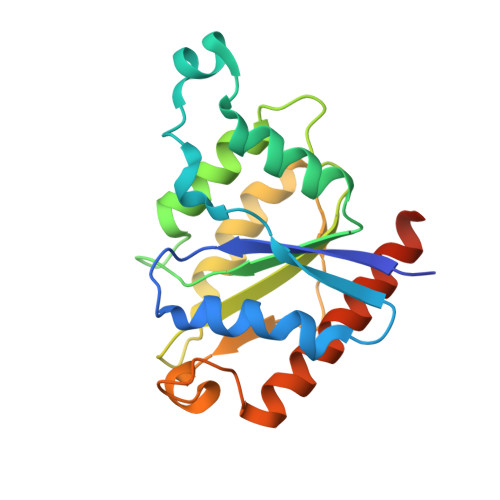
3S2Y - PubMed Abstract:
Environmental protection through biological mechanisms that aid in the reductive immobilization of toxic metals (e.g., chromate and uranyl) has been identified to involve specific NADH-dependent flavoproteins that promote cell viability. To understand the enzyme mechanisms responsible for metal reduction, the enzyme kinetics of a putative chromate reductase from Gluconacetobacter hansenii (Gh-ChrR) was measured and the crystal structure of the protein determined at 2.25 Å resolution. Gh-ChrR catalyzes the NADH-dependent reduction of chromate, ferricyanide, and uranyl anions under aerobic conditions. Kinetic measurements indicate that NADH acts as a substrate inhibitor; catalysis requires chromate binding prior to NADH association. The crystal structure of Gh-ChrR shows the protein is a homotetramer with one bound flavin mononucleotide (FMN) per subunit. A bound anion is visualized proximal to the FMN at the interface between adjacent subunits within a cationic pocket, which is positioned at an optimal distance for hydride transfer. Site-directed substitutions of residues proposed to involve in both NADH and metal anion binding (N85A or R101A) result in 90-95% reductions in enzyme efficiencies for NADH-dependent chromate reduction. In comparison site-directed substitution of a residue (S118A) participating in the coordination of FMN in the active site results in only modest (50%) reductions in catalytic efficiencies, consistent with the presence of a multitude of side chains that position the FMN in the active site. The proposed proximity relationships between metal anion binding site and enzyme cofactors is discussed in terms of rational design principles for the use of enzymes in chromate and uranyl bioremediation.
- Biological Sciences Division, Pacific Northwest National Laboratory, Richland, Washington, United States of America. hongjunj@mir.wustl.edu
Organizational Affiliation: