3S0H | pdb_00003s0h

The crystal structure of the periplasmic domain of Helicobacter pylori MotB (residues 90-256).


Experimental Data Snapshot

  • Method: X-RAY DIFFRACTION
  • Resolution: 2.10 Å
  • R-Value Free: 
    0.236 (Depositor) 
  • R-Value Work: 
    0.181 (Depositor) 
  • R-Value Observed: 
    0.184 (Depositor) 

wwPDB Validation 3D Report Full Report

Validation slider image for 3S0H

This is version 1.1 of the entry. See complete history

Literature

Role of the MotB linker in the assembly and activation of the bacterial flagellar motor.

O'Neill, J.Xie, M.Hijnen, M.Roujeinikova, A.

(2011) Acta Crystallogr D Biol Crystallogr 67: 1009-1016

  • DOI: https://doi.org/10.1107/S0907444911041102
  • Primary Citation Related Structures: 
    3S02, 3S03, 3S06, 3S0H, 3S0W, 3S0Y

  • PubMed Abstract: 

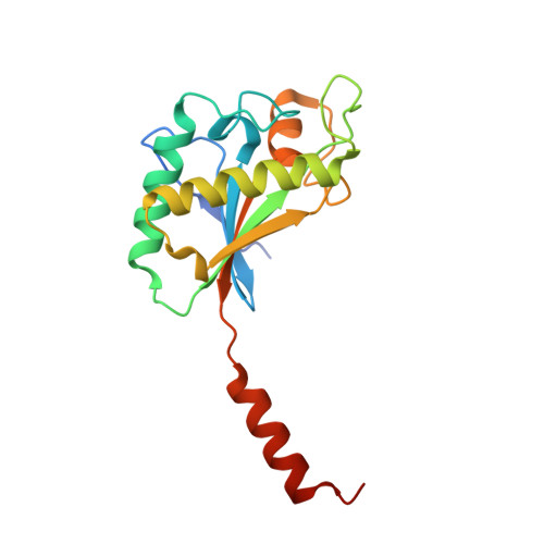
    Bacterial flagella are driven by an ion influx through the peptidoglycan (PG)-tethered MotA/MotB stator. Stator precomplexes assemble in the membrane and remain inactive until they incorporate into the motor, upon which MotA/MotB changes conformation. The nature of this change and the mechanism of inhibition of the PG-binding and ion-conducting activities of the precomplexes are unknown. Here, the structural analysis of a series of N-terminally truncated MotB fragments is presented, the mechanism of inhibition by the linker is identified and the structural basis for the formation of the PG-binding-competent open-channel MotA/MotB conformation via a mechanism that entails linker unfolding and rotational displacement of MotB transmembrane helices is uncovered.


  • Organizational Affiliation
    • Manchester Interdisciplinary Biocentre, University of Manchester, 131 Princess Street, Manchester M1 7DN, England.

Macromolecule Content 

  • Total Structure Weight: 79.53 kDa 
  • Atom Count: 6,059 
  • Modeled Residue Count: 650 
  • Deposited Residue Count: 692 
  • Unique protein chains: 1

Macromolecules

Find similar proteins by:|  3D Structure
Entity ID: 1
MoleculeChains  Sequence LengthOrganismDetailsImage
Motility protein B
A, B, C, D
173Helicobacter pyloriMutation(s): 0 
Gene Names: motBHP_0816
UniProt
Find proteins for P56427 (Helicobacter pylori (strain ATCC 700392 / 26695))
Explore P56427 
Go to UniProtKB:  P56427
Entity Groups
Sequence Clusters30% Identity50% Identity70% Identity90% Identity95% Identity100% Identity
UniProt GroupP56427
Sequence Annotations
Expand
Reference Sequence

Small Molecules

Ligands 1 Unique
IDChains Name / Formula / InChI Key2D Diagram3D Interactions
SO4

Query on SO4


Download:Ideal Coordinates CCD File
E [auth A]
F [auth A]
G [auth B]
H [auth B]
I [auth B]
E [auth A],
F [auth A],
G [auth B],
H [auth B],
I [auth B],
J [auth C],
K [auth C],
L [auth C],
M [auth D]
SULFATE ION
O4 S
QAOWNCQODCNURD-UHFFFAOYSA-L

Experimental Data & Validation

Experimental Data

  • Method: X-RAY DIFFRACTION
  • Resolution: 2.10 Å
  • R-Value Free:  0.236 (Depositor) 
  • R-Value Work:  0.181 (Depositor) 
  • R-Value Observed: 0.184 (Depositor) 
Space Group: P 43
Unit Cell:
Length ( Å )Angle ( ˚ )
a = 72.972α = 90
b = 72.972β = 90
c = 127.178γ = 90
Software Package:
Software NamePurpose
SCALAdata scaling
REFMACrefinement
PDB_EXTRACTdata extraction
PHASERphasing

Structure Validation

View Full Validation Report


Entry History 

Deposition Data

Revision History  (Full details and data files)

  • Version 1.0: 2012-03-14
    Type: Initial release
  • Version 1.1: 2024-02-28
    Changes: Data collection, Database references, Derived calculations