3RX6

Crystal structure of Polarity Suppression protein from Enterobacteria phage P4


Experimental Data Snapshot

  • Method: X-RAY DIFFRACTION
  • Resolution: 2.04 Å
  • R-Value Free: 0.219 
  • R-Value Work: 0.192 
  • R-Value Observed: 0.195 

wwPDB Validation   3D Report Full Report


This is version 1.3 of the entry. See complete history


Literature

The first structure of polarity suppression protein, Psu from enterobacteria phage P4, reveals a novel fold and a knotted dimer

Banerjee, R.Nath, S.Ranjan, A.Khamrui, S.Pani, B.Sen, R.Sen, U.

(2012) J Biol Chem 287: 44667-44675

  • DOI: https://doi.org/10.1074/jbc.M112.423202
  • Primary Citation of Related Structures:  
    3RX6, 4DVD

  • PubMed Abstract: 

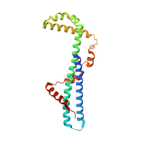
    Psu is a capsid decoration protein of bacteriophage P4 and acts as an antiterminator of Rho-dependent transcription termination in bacteria. So far, no structures have been reported for the Psu protein or its homologues. Here, we report the first structure of Psu solved by the Hg(2+) single wavelength anomalous dispersion method, which reveals that Psu exists as a knotted homodimer and is first of its kind in nature. Each monomer of Psu attains a novel fold around a tight coiled-coil motif. CD spectroscopy and the structure of an engineered disulfide-bridged Psu derivative reveal that the protein folds reversibly and reassembles by itself into the knotted dimeric conformation without the requirement of any chaperone. This structure would help to explain the functional properties of the protein and can be used as a template to design a minimal peptide fragment that can be used as a drug against Rho-dependent transcription termination in bacteria.


  • Organizational Affiliation

    Crystallography and Molecular Biology Division, Saha Institute of Nuclear Physics, 1/AF, Bidhannagar, Kolkata 700064, India.


Macromolecules
Find similar proteins by:  (by identity cutoff)  |  3D Structure
Entity ID: 1
MoleculeChains Sequence LengthOrganismDetailsImage
Polarity suppression protein190Enterobacteria phage P4Mutation(s): 0 
Gene Names: psu
UniProt
Find proteins for P05460 (Enterobacteria phage P4)
Explore P05460 
Go to UniProtKB:  P05460
Entity Groups  
Sequence Clusters30% Identity50% Identity70% Identity90% Identity95% Identity100% Identity
UniProt GroupP05460
Sequence Annotations
Expand
  • Reference Sequence
Experimental Data & Validation

Experimental Data

  • Method: X-RAY DIFFRACTION
  • Resolution: 2.04 Å
  • R-Value Free: 0.219 
  • R-Value Work: 0.192 
  • R-Value Observed: 0.195 
  • Space Group: I 4 2 2
Unit Cell:
Length ( Å )Angle ( ˚ )
a = 148.75α = 90
b = 148.75β = 90
c = 63.37γ = 90
Software Package:
Software NamePurpose
MAR345dtbdata collection
SOLVEphasing
PHENIXrefinement
iMOSFLMdata reduction
SCALAdata scaling

Structure Validation

View Full Validation Report


Entry History 

Deposition Data

Revision History  (Full details and data files)

  • Version 1.0: 2012-07-25
    Type: Initial release
  • Version 1.1: 2012-11-21
    Changes: Database references
  • Version 1.2: 2013-06-19
    Changes: Database references
  • Version 1.3: 2024-03-20
    Changes: Data collection, Database references, Derived calculations