3RUI

Crystal structure of Atg7C-Atg8 complex


Experimental Data Snapshot

  • Method: X-RAY DIFFRACTION
  • Resolution: 1.91 Å
  • R-Value Free: 0.218 
  • R-Value Work: 0.192 
  • R-Value Observed: 0.193 

wwPDB Validation   3D Report Full Report


This is version 1.1 of the entry. See complete history


Literature

Insights into noncanonical E1 enzyme activation from the structure of autophagic E1 Atg7 with Atg8.

Hong, S.B.Kim, B.W.Lee, K.E.Kim, S.W.Jeon, H.Kim, J.Song, H.K.

(2011) Nat Struct Mol Biol 18: 1323-1330

  • DOI: https://doi.org/10.1038/nsmb.2165
  • Primary Citation of Related Structures:  
    3RUI, 3RUJ

  • PubMed Abstract: 

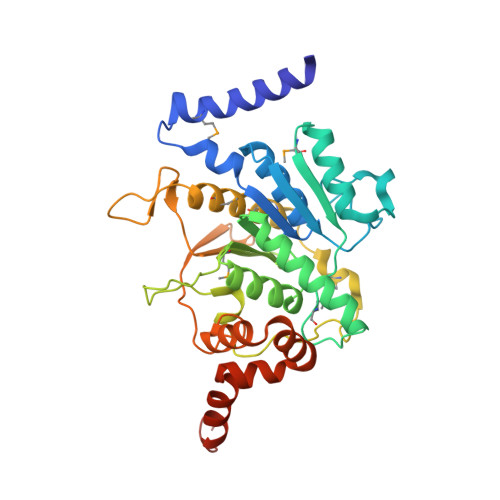
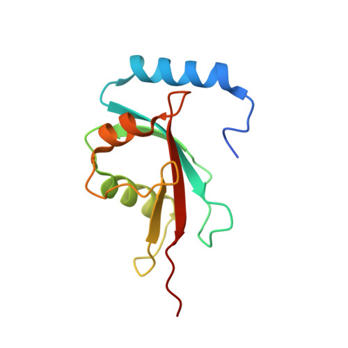
    Autophagy is the degradation of cellular organelles via the lysosomal pathway. The autophagic ubiquitin-like (Ubl) molecule Atg8 is activated by the E1-like enzyme Atg7. As this noncanonical E1 enzyme's domain organization is unique among Ubl-activating E1 enzymes, the structural basis for its interactions with Atg8 and partner E2 enzymes remains obscure. Here we present the structure of the N-terminal domain of Atg7, revealing a unique protein fold and interactions with both autophagic E2 enzymes Atg3 and Atg10. The structure of the C-terminal domain of Atg7 in complex with Atg8 shows the mode of dimerization and mechanism of recognition of Atg8. Notably, the catalytic cysteine residue in Atg7 is positioned close to the C-terminal glycine of Atg8, its target for thioester formation, potentially eliminating the need for large conformational rearrangements characteristic of other E1s.


  • Organizational Affiliation

    School of Life Sciences and Biotechnology, Korea University, Seoul, South Korea.


Macromolecules
Find similar proteins by:  (by identity cutoff)  |  3D Structure
Entity ID: 1
MoleculeChains Sequence LengthOrganismDetailsImage
Ubiquitin-like modifier-activating enzyme ATG7340Saccharomyces cerevisiae S288CMutation(s): 1 
Gene Names: ATG7APG7CVT2YHR171W
UniProt
Find proteins for P38862 (Saccharomyces cerevisiae (strain ATCC 204508 / S288c))
Explore P38862 
Go to UniProtKB:  P38862
Entity Groups  
Sequence Clusters30% Identity50% Identity70% Identity90% Identity95% Identity100% Identity
UniProt GroupP38862
Sequence Annotations
Expand
  • Reference Sequence
Find similar proteins by:  (by identity cutoff)  |  3D Structure
Entity ID: 2
MoleculeChains Sequence LengthOrganismDetailsImage
Autophagy-related protein 8118Saccharomyces cerevisiae S288CMutation(s): 0 
Gene Names: ATG8APG8AUT7CVT5YBL078CYBL0732
UniProt
Find proteins for P38182 (Saccharomyces cerevisiae (strain ATCC 204508 / S288c))
Explore P38182 
Go to UniProtKB:  P38182
Entity Groups  
Sequence Clusters30% Identity50% Identity70% Identity90% Identity95% Identity100% Identity
UniProt GroupP38182
Sequence Annotations
Expand
  • Reference Sequence
Small Molecules
Ligands 1 Unique
IDChains Name / Formula / InChI Key2D Diagram3D Interactions
ZN
Query on ZN

Download Ideal Coordinates CCD File 
C [auth A]ZINC ION
Zn
PTFCDOFLOPIGGS-UHFFFAOYSA-N
Modified Residues  1 Unique
IDChains TypeFormula2D DiagramParent
MSE
Query on MSE
A
L-PEPTIDE LINKINGC5 H11 N O2 SeMET
Experimental Data & Validation

Experimental Data

  • Method: X-RAY DIFFRACTION
  • Resolution: 1.91 Å
  • R-Value Free: 0.218 
  • R-Value Work: 0.192 
  • R-Value Observed: 0.193 
  • Space Group: P 43 21 2
Unit Cell:
Length ( Å )Angle ( ˚ )
a = 71.326α = 90
b = 71.326β = 90
c = 220.916γ = 90
Software Package:
Software NamePurpose
HKL-2000data collection
SOLVEphasing
PHENIXrefinement
HKL-2000data reduction
HKL-2000data scaling

Structure Validation

View Full Validation Report


Entry History 

Deposition Data

Revision History  (Full details and data files)

  • Version 1.0: 2011-11-23
    Type: Initial release
  • Version 1.1: 2013-07-03
    Changes: Database references