3RSP | pdb_00003rsp

STRUCTURE OF THE P93G VARIANT OF RIBONUCLEASE A


Experimental Data Snapshot

  • Method: X-RAY DIFFRACTION
  • Resolution: 1.70 Å
  • R-Value Work: 
    0.170 (Depositor), 0.159 (DCC) 

Starting Model: experimental
View more details

wwPDB Validation 3D Report Full Report

Validation slider image for 3RSP

This is version 1.5 of the entry. See complete history

Literature

Structure and stability of the P93G variant of ribonuclease A.

Schultz, L.W.Hargraves, S.R.Klink, T.A.Raines, R.T.

(1998) Protein Sci 7: 1620-1625

  • DOI: https://doi.org/10.1002/pro.5560070716
  • Primary Citation Related Structures: 
    3RSP

  • PubMed Abstract: 

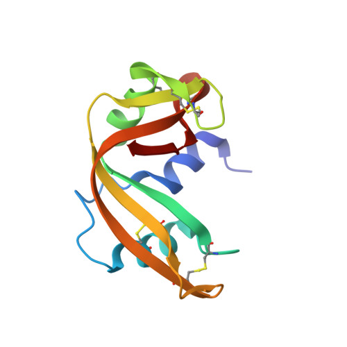
    The peptide bonds preceding Pro 93 and Pro 114 of bovine pancreatic ribonuclease A (RNase A) are in the cis conformation. The trans-to-cis isomerization of these bonds had been indicted as the slow step during protein folding. Here, site-directed mutagenesis was used to replace Pro 93 or Pro 114 with a glycine residue, and the crystalline structure of the P93G variant was determined by X-ray diffraction analysis to a resolution of 1.7 A. This structure is essentially identical to that of the wild-type protein, except for the 91-94 beta-turn containing the substitution. In the wild-type protein, the beta-turn is of type VIa. In the P93G variant, this turn is of type II with the peptide bond preceding Gly 93 being trans. The thermal stabilities of the P93G and P114G variants were assessed by differential scanning calorimetry and thermal denaturation experiments monitored by ultraviolet spectroscopy. The value of delta deltaGm which reports on the stability lost in the variants, is 1.5-fold greater for the P114G variant than for the P93G variant. The greater stability of the P93G variant is likely due to the relatively facile accommodation of residues 91-94 in a type II turn, which has a preference for a glycine residue in its i + 2 position.


  • Organizational Affiliation
    • Department of Biochemistry, University of Wisconsin-Madison, 53706-1569, USA.

Macromolecule Content 

  • Total Structure Weight: 13.74 kDa 
  • Atom Count: 1,108 
  • Modeled Residue Count: 124 
  • Deposited Residue Count: 124 
  • Unique protein chains: 1

Macromolecules

Find similar proteins by:|  3D Structure
Entity ID: 1
MoleculeChains  Sequence LengthOrganismDetailsImage
RIBONUCLEASE A124Bos taurusMutation(s): 1 
EC: 3.1.27.5 (PDB Primary Data), 4.6.1.18 (UniProt)
UniProt
Find proteins for P61823 (Bos taurus)
Explore P61823 
Go to UniProtKB:  P61823
Entity Groups
Sequence Clusters30% Identity50% Identity70% Identity90% Identity95% Identity100% Identity
UniProt GroupP61823
Sequence Annotations
Expand
Reference Sequence

Experimental Data & Validation

Experimental Data

  • Method: X-RAY DIFFRACTION
  • Resolution: 1.70 Å
  • R-Value Work:  0.170 (Depositor), 0.159 (DCC) 
Space Group: P 32 2 1
Unit Cell:
Length ( Å )Angle ( ˚ )
a = 65.79α = 90
b = 65.79β = 90
c = 66.12γ = 120
Software Package:
Software NamePurpose
XDSdata scaling
XCALIBREdata reduction
AMoREphasing
TNTrefinement
XDSdata reduction
XCALIBREdata scaling

Structure Validation

View Full Validation Report


Entry History 

Deposition Data

Revision History  (Full details and data files)

  • Version 1.0: 1998-04-22
    Type: Initial release
  • Version 1.1: 2008-03-25
    Changes: Version format compliance
  • Version 1.2: 2011-07-13
    Changes: Version format compliance
  • Version 1.3: 2021-11-03
    Changes: Database references, Derived calculations
  • Version 1.4: 2023-08-09
    Changes: Refinement description
  • Version 1.5: 2024-10-30
    Changes: Data collection, Structure summary