3RS1 | pdb_00003rs1

Mouse C-type lectin-related protein Clrg


Experimental Data Snapshot

  • Method: X-RAY DIFFRACTION
  • Resolution: 1.94 Å
  • R-Value Free: 
    0.204 (DCC) 
  • R-Value Work: 
    0.185 (Depositor), 0.193 (DCC) 
  • R-Value Observed: 
    0.186 (Depositor) 

Starting Model: experimental
View more details

wwPDB Validation 3D Report Full Report

Validation slider image for 3RS1

This is version 1.4 of the entry. See complete history

Literature

Mouse Clr-g, a Ligand for NK Cell Activation Receptor NKR-P1F: Crystal Structure and Biophysical Properties.

Skalova, T.Kotynkova, K.Duskova, J.Hasek, J.Kovai, T.Kolenko, P.Novak, P.Man, P.Hanc, P.Vanek, O.Bezouska, K.Dohnalek, J.

(2012) J Immunol 189: 4881-4889

  • DOI: https://doi.org/10.4049/jimmunol.1200880
  • Primary Citation Related Structures: 
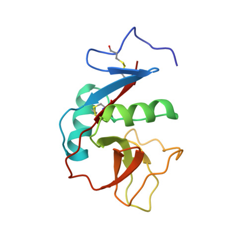
    3RS1

  • PubMed Abstract: 

    Interactions between C-type lectin-like NK cell receptors and their protein ligands form one of the key recognition mechanisms of the innate immune system that is involved in the elimination of cells that have been malignantly transformed, virally infected, or stressed by chemotherapy or other factors. We determined an x-ray structure for the extracellular domain of mouse C-type lectin related (Clr) protein g, a ligand for the activation receptor NKR-P1F. Clr-g forms dimers in the crystal structure resembling those of human CD69. This newly reported structure, together with the previously determined structure of mouse receptor NKR-P1A, allowed the modeling and calculations of electrostatic profiles for other closely related receptors and ligands. Despite the high similarity among Clr-g, Clr-b, and human CD69, these molecules have fundamentally different electrostatics, with distinct polarization of Clr-g. The electrostatic profile of NKR-P1F is complementary to that of Clr-g, which suggests a plausible interaction mechanism based on contacts between surface sites of opposite potential.


  • Organizational Affiliation
    • Institute of Macromolecular Chemistry, Academy of Sciences of the Czech Republic, vvi, 16206 Praha 6, Czech Republic. t.skalova@gmail.com

Macromolecule Content 

  • Total Structure Weight: 28.89 kDa 
  • Atom Count: 2,311 
  • Modeled Residue Count: 244 
  • Deposited Residue Count: 244 
  • Unique protein chains: 1

Macromolecules

Find similar proteins by:|  3D Structure
Entity ID: 1
MoleculeChains  Sequence LengthOrganismDetailsImage
C-type lectin domain family 2 member I
A, B
122Mus musculusMutation(s): 2 
Gene Names: Clec2iClrgDcl1Ocilrp2
UniProt
Find proteins for Q9WVF9 (Mus musculus)
Explore Q9WVF9 
Go to UniProtKB:  Q9WVF9
Entity Groups
Sequence Clusters30% Identity50% Identity70% Identity90% Identity95% Identity100% Identity
UniProt GroupQ9WVF9
Sequence Annotations
Expand
Reference Sequence

Small Molecules

Ligands 1 Unique
IDChains Name / Formula / InChI Key2D Diagram3D Interactions
CL

Query on CL


Download:Ideal Coordinates CCD File
C [auth A]CHLORIDE ION
Cl
VEXZGXHMUGYJMC-UHFFFAOYSA-M

Experimental Data & Validation

Experimental Data

  • Method: X-RAY DIFFRACTION
  • Resolution: 1.94 Å
  • R-Value Free:  0.204 (DCC) 
  • R-Value Work:  0.185 (Depositor), 0.193 (DCC) 
  • R-Value Observed: 0.186 (Depositor) 
Space Group: P 21 21 2
Unit Cell:
Length ( Å )Angle ( ˚ )
a = 118.31α = 90
b = 61.38β = 90
c = 32.36γ = 90
Software Package:
Software NamePurpose
MxCuBEdata collection
BALBESphasing
REFMACrefinement
HKL-2000data reduction
HKL-2000data scaling

Structure Validation

View Full Validation Report


Entry History 

Deposition Data

Revision History  (Full details and data files)

  • Version 1.0: 2012-05-02
    Type: Initial release
  • Version 1.1: 2012-11-14
    Changes: Database references
  • Version 1.2: 2012-12-19
    Changes: Database references
  • Version 1.3: 2023-09-13
    Changes: Data collection, Database references, Derived calculations, Refinement description
  • Version 1.4: 2024-11-06
    Changes: Structure summary