3RQR

Crystal structure of the RYR domain of the rabbit ryanodine receptor


Experimental Data Snapshot

  • Method: X-RAY DIFFRACTION
  • Resolution: 2.16 Å
  • R-Value Free: 0.258 
  • R-Value Work: 0.231 
  • R-Value Observed: 0.238 

wwPDB Validation   3D Report Full Report


This is version 1.4 of the entry. See complete history


Literature

Structural determination of the phosphorylation domain of the ryanodine receptor.

Sharma, P.Ishiyama, N.Nair, U.Li, W.Dong, A.Miyake, T.Wilson, A.Ryan, T.Maclennan, D.H.Kislinger, T.Ikura, M.Dhe-Paganon, S.Gramolini, A.O.

(2012) FEBS J 279: 3952-3964

  • DOI: https://doi.org/10.1111/j.1742-4658.2012.08755.x
  • Primary Citation of Related Structures:  
    3RQR

  • PubMed Abstract: 

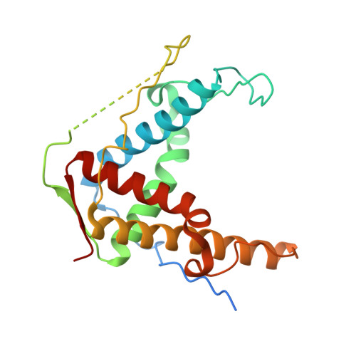
    The ryanodine receptor (RyR) is a large, homotetrameric sarcoplasmic reticulum membrane protein that is essential for Ca(2+) cycling in both skeletal and cardiac muscle. Genetic mutations in RyR1 are associated with severe conditions including malignant hyperthermia (MH) and central core disease. One phosphorylation site (Ser 2843) has been identified in a segment of RyR1 flanked by two RyR motifs, which are found exclusively in all RyR isoforms as closely associated tandem (or paired) motifs, and are named after the protein itself. These motifs also contain six known MH mutations. In this study, we designed, expressed and purified the tandem RyR motifs, and show that this domain contains a putative binding site for the Ca(2+)/calmodulin-dependent protein kinase β isoform. We present a 2.2 Å resolution crystal structure of the RyR domain revealing a two-fold, symmetric, extended four-helix bundle stabilized by a β sheet. Using mathematical modelling, we fit our crystal structure within a tetrameric electron microscopy (EM) structure of native RyR1, and propose that this domain is localized in the RyR clamp region, which is absent in its cousin protein inositol 1,4,5-trisphosphate receptor.


  • Organizational Affiliation

    Department of Physiology, University of Toronto, Toronto, Ontario, Canada.


Macromolecules
Find similar proteins by:  (by identity cutoff)  |  3D Structure
Entity ID: 1
MoleculeChains Sequence LengthOrganismDetailsImage
Ryanodine receptor 1227Oryctolagus cuniculusMutation(s): 0 
Gene Names: RYR1
UniProt
Find proteins for P11716 (Oryctolagus cuniculus)
Explore P11716 
Go to UniProtKB:  P11716
Entity Groups  
Sequence Clusters30% Identity50% Identity70% Identity90% Identity95% Identity100% Identity
UniProt GroupP11716
Sequence Annotations
Expand
  • Reference Sequence

Find similar proteins by:  Sequence   |   3D Structure  

Entity ID: 2
MoleculeChains Sequence LengthOrganismDetailsImage
(UNK)(UNK)(UNK)(UNK)B [auth U]4Oryctolagus cuniculusMutation(s): 0 
Sequence Annotations
Expand
  • Reference Sequence
Experimental Data & Validation

Experimental Data

  • Method: X-RAY DIFFRACTION
  • Resolution: 2.16 Å
  • R-Value Free: 0.258 
  • R-Value Work: 0.231 
  • R-Value Observed: 0.238 
  • Space Group: P 43 21 2
Unit Cell:
Length ( Å )Angle ( ˚ )
a = 68.77α = 90
b = 68.77β = 90
c = 91.38γ = 90
Software Package:
Software NamePurpose
HKL-3000data collection
SHELXSphasing
SOLVEphasing
RESOLVEmodel building
BUSTERrefinement
Cootmodel building
HKL-3000data reduction
HKL-3000data scaling
RESOLVEphasing

Structure Validation

View Full Validation Report


Entry History 

Revision History  (Full details and data files)

  • Version 1.0: 2011-06-22
    Type: Initial release
  • Version 1.1: 2011-07-13
    Changes: Version format compliance
  • Version 1.2: 2012-09-05
    Changes: Database references
  • Version 1.3: 2012-10-17
    Changes: Database references
  • Version 1.4: 2024-02-28
    Changes: Data collection, Database references