3RPI | pdb_00003rpi

Crystal Structure of Fab from 3BNC60, Highly Potent anti-HIV Antibody


Experimental Data Snapshot

  • Method: X-RAY DIFFRACTION
  • Resolution: 2.65 Å
  • R-Value Free: 
    0.258 (Depositor), 0.260 (DCC) 
  • R-Value Work: 
    0.208 (Depositor), 0.204 (DCC) 
  • R-Value Observed: 
    0.210 (Depositor) 

wwPDB Validation 3D Report Full Report

Validation slider image for 3RPI

Ligand Structure Quality Assessment 


This is version 1.4 of the entry. See complete history

Literature

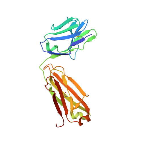
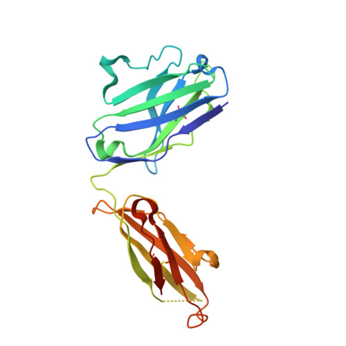
Sequence and structural convergence of broad and potent HIV antibodies that mimic CD4 binding.

Scheid, J.F.Mouquet, H.Ueberheide, B.Diskin, R.Klein, F.Oliveira, T.Y.Pietzsch, J.Fenyo, D.Abadir, A.Velinzon, K.Hurley, A.Myung, S.Boulad, F.Poignard, P.Burton, D.R.Pereyra, F.Ho, D.D.Walker, B.D.Seaman, M.S.Bjorkman, P.J.Chait, B.T.Nussenzweig, M.C.

(2011) Science 333: 1633-1637

  • DOI: https://doi.org/10.1126/science.1207227
  • Primary Citation Related Structures: 
    3RPI

  • PubMed Abstract: 

    Passive transfer of broadly neutralizing HIV antibodies can prevent infection, which suggests that vaccines that elicit such antibodies would be protective. Thus far, however, few broadly neutralizing HIV antibodies that occur naturally have been characterized. To determine whether these antibodies are part of a larger group of related molecules, we cloned 576 new HIV antibodies from four unrelated individuals. All four individuals produced expanded clones of potent broadly neutralizing CD4-binding-site antibodies that mimic binding to CD4. Despite extensive hypermutation, the new antibodies shared a consensus sequence of 68 immunoglobulin H (IgH) chain amino acids and arise independently from two related IgH genes. Comparison of the crystal structure of one of the antibodies to the broadly neutralizing antibody VRC01 revealed conservation of the contacts to the HIV spike.


  • Organizational Affiliation
    • Laboratory of Molecular Immunology, The Rockefeller University, New York, NY 10065, USA.

Macromolecule Content 

  • Total Structure Weight: 96.41 kDa 
  • Atom Count: 6,650 
  • Modeled Residue Count: 830 
  • Deposited Residue Count: 870 
  • Unique protein chains: 2

Macromolecules

Find similar proteins by:|  3D Structure
Entity ID: 1
MoleculeChains  Sequence LengthOrganismDetailsImage
Light chain from highly potent anti-HIV neutralizing antibodyA [auth L],
D [auth B]
206Homo sapiensMutation(s): 0 
Find similar proteins by:|  3D Structure
Entity ID: 2
MoleculeChains  Sequence LengthOrganismDetailsImage
Heavy chain from highly potent anti-HIV neutralizing antibodyB [auth H],
C [auth A]
229Homo sapiensMutation(s): 0 

Experimental Data & Validation

Experimental Data

  • Method: X-RAY DIFFRACTION
  • Resolution: 2.65 Å
  • R-Value Free:  0.258 (Depositor), 0.260 (DCC) 
  • R-Value Work:  0.208 (Depositor), 0.204 (DCC) 
  • R-Value Observed: 0.210 (Depositor) 
Space Group: P 1 21 1
Unit Cell:
Length ( Å )Angle ( ˚ )
a = 63.62α = 90
b = 155.68β = 110.4
c = 74.76γ = 90
Software Package:
Software NamePurpose
Web-Icedata collection
PHASERphasing
PHENIXrefinement
XDSdata reduction
XDSdata scaling

Structure Validation

View Full Validation Report


Ligand Structure Quality Assessment 


Entry History 

Deposition Data

Revision History  (Full details and data files)

  • Version 1.0: 2011-07-20
    Type: Initial release
  • Version 1.1: 2011-08-03
    Changes: Database references, Structure summary
  • Version 1.2: 2011-09-28
    Changes: Database references
  • Version 1.3: 2020-07-29
    Type: Remediation
    Reason: Carbohydrate remediation
    Changes: Data collection, Database references, Derived calculations, Refinement description, Structure summary
  • Version 1.4: 2024-11-27
    Changes: Data collection, Database references, Structure summary