


Crystal structure of C105S mutant of Mycobacterium tuberculosis methionine aminopeptidase
Reddi, R., Kishor, C., Addlagatta, A.To be published.
Experimental Data Snapshot
Starting Model: experimental
View more details
wwPDB Validation 3D Report Full Report

Entity ID: 1 | |||||
|---|---|---|---|---|---|
| Molecule | Chains | Sequence Length | Organism | Details | Image |
| Methionine aminopeptidase | 291 | Mycobacterium tuberculosis | Mutation(s): 1 Gene Names: map, mapB, MetAP, MT2929, MTV003.07c, Rv2861c EC: 3.4.11.18 | 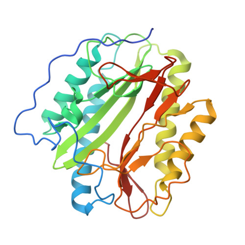 | |
UniProt | |||||
Entity Groups | |||||
| Sequence Clusters | 30% Identity50% Identity70% Identity90% Identity95% Identity100% Identity | ||||
| UniProt Group | P9WK19 | ||||
Sequence AnnotationsExpand | |||||
Reference Sequence | |||||
| Ligands 3 Unique | |||||
|---|---|---|---|---|---|
| ID | Chains | Name / Formula / InChI Key | 2D Diagram | 3D Interactions | |
| BME Download:Ideal Coordinates CCD File | E [auth A] | BETA-MERCAPTOETHANOL C2 H6 O S DGVVWUTYPXICAM-UHFFFAOYSA-N |  | ||
| CO Download:Ideal Coordinates CCD File | B [auth A], C [auth A] | COBALT (II) ION Co XLJKHNWPARRRJB-UHFFFAOYSA-N |  | ||
| K Download:Ideal Coordinates CCD File | D [auth A] | POTASSIUM ION K NPYPAHLBTDXSSS-UHFFFAOYSA-N |  | ||
| Length ( Å ) | Angle ( ˚ ) |
|---|---|
| a = 49.22 | α = 90 |
| b = 48.66 | β = 95.16 |
| c = 55.72 | γ = 90 |
| Software Name | Purpose |
|---|---|
| ADSC | data collection |
| MOLREP | phasing |
| REFMAC | refinement |
| MOSFLM | data reduction |
| SCALA | data scaling |


