3ROF

Crystal Structure of the S. aureus Protein Tyrosine Phosphatase PtpA


Experimental Data Snapshot

  • Method: X-RAY DIFFRACTION
  • Resolution: 1.03 Å
  • R-Value Free: 0.169 
  • R-Value Work: 0.149 
  • R-Value Observed: 0.150 

wwPDB Validation   3D Report Full Report


This is version 1.3 of the entry. See complete history


Literature

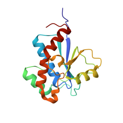
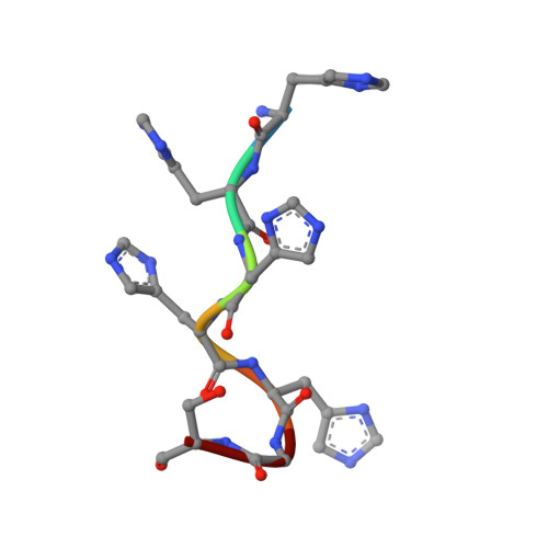
Structure and substrate recognition of the Staphylococcus aureus protein tyrosine phosphatase PtpA.

Vega, C.Chou, S.Engel, K.Harrell, M.E.Rajagopal, L.Grundner, C.

(2011) J Mol Biol 413: 24-31

  • DOI: https://doi.org/10.1016/j.jmb.2011.08.015
  • Primary Citation of Related Structures:  
    3ROF

  • PubMed Abstract: 

    Phosphosignaling through pSer/pThr/pTyr is emerging as a common signaling mechanism in prokaryotes. The human pathogen Staphylococcus aureus produces two low-molecular-weight protein tyrosine phosphatases (PTPs), PtpA and PtpB, with unknown functions. To provide the structural context for understanding PtpA function and substrate recognition, establish PtpA's structural relations within the PTP family, and provide a framework for the design of specific inhibitors, we solved the crystal structure of PtpA at 1 Å resolution. While PtpA adopts the common, conserved PTP fold and shows close overall similarity to eukaryotic PTPs, several features in the active site and surface organization are unique and can be explored to design selective inhibitors. A peptide bound in the active site mimics a phosphotyrosine substrate, affords insight into substrate recognition, and provides a testable substrate prediction. Genetic deletion of ptpA or ptpB does not affect in vitro growth or cell wall integrity, raising the possibility that PtpA and PtpB have specialized functions during infection.


  • Organizational Affiliation

    Seattle Biomedical Research Institute, Seattle, WA 98109, USA.


Macromolecules
Find similar proteins by:  (by identity cutoff)  |  3D Structure
Entity ID: 1
MoleculeChains Sequence LengthOrganismDetailsImage
Low molecular weight protein-tyrosine-phosphatase ptpA158Staphylococcus aureusMutation(s): 0 
Gene Names: ptpA
EC: 3.1.3.48
UniProt
Find proteins for P0C5D2 (Staphylococcus aureus)
Explore P0C5D2 
Go to UniProtKB:  P0C5D2
Entity Groups  
Sequence Clusters30% Identity50% Identity70% Identity90% Identity95% Identity100% Identity
UniProt GroupP0C5D2
Sequence Annotations
Expand
  • Reference Sequence

Find similar proteins by:  Sequence   |   3D Structure  

Entity ID: 2
MoleculeChains Sequence LengthOrganismDetailsImage
Expression tag cleaved from protein-tyrosine-phosphatase ptpA7Staphylococcus aureusMutation(s): 0 
Gene Names: ptpA
Sequence Annotations
Expand
  • Reference Sequence
Experimental Data & Validation

Experimental Data

  • Method: X-RAY DIFFRACTION
  • Resolution: 1.03 Å
  • R-Value Free: 0.169 
  • R-Value Work: 0.149 
  • R-Value Observed: 0.150 
  • Space Group: P 1 21 1
Unit Cell:
Length ( Å )Angle ( ˚ )
a = 34.81α = 90
b = 35.1β = 91.05
c = 65.51γ = 90
Software Package:
Software NamePurpose
DENZOdata reduction
SCALEPACKdata scaling
PHENIXrefinement
PDB_EXTRACTdata extraction
ADSCdata collection
XDSdata reduction
XDSdata scaling
MOLREPphasing

Structure Validation

View Full Validation Report


Entry History 

Deposition Data

Revision History  (Full details and data files)

  • Version 1.0: 2011-09-07
    Type: Initial release
  • Version 1.1: 2012-05-23
    Changes: Database references
  • Version 1.2: 2017-11-08
    Changes: Advisory, Refinement description
  • Version 1.3: 2024-02-28
    Changes: Advisory, Data collection, Database references, Derived calculations